Message Board

Respected readers, authors and reviewers, you can add comments to this page on any questions about the contribution, review,        editing and publication of this journal. We will give you an answer as soon as possible. Thank you for your support!

Name
E-mail
Phone
Title
Content
Verification Code
Turn off MathJax
Article Contents

ZHANG Tingyan, LI Tingting, LUO Yunchun, YUAN Xiaofeng, ZHANG Chuan, HAO Kai, BIAN Jun. Exploring the mechanism of tanshinone ⅡA in ameliorating acetaminophen-induced liver injury based on network pharmacology and molecular docking techniques[J]. Journal of Pharmaceutical Practice and Service. doi: 10.12206/j.issn.2097-2024.202412022
Citation: ZHANG Tingyan, LI Tingting, LUO Yunchun, YUAN Xiaofeng, ZHANG Chuan, HAO Kai, BIAN Jun. Exploring the mechanism of tanshinone ⅡA in ameliorating acetaminophen-induced liver injury based on network pharmacology and molecular docking techniques[J]. Journal of Pharmaceutical Practice and Service. doi: 10.12206/j.issn.2097-2024.202412022

Exploring the mechanism of tanshinone ⅡA in ameliorating acetaminophen-induced liver injury based on network pharmacology and molecular docking techniques

doi: 10.12206/j.issn.2097-2024.202412022
  • Received Date: 2024-12-09
  • Accepted Date: 2026-05-27
  • Rev Recd Date: 2025-05-26
  •   Objective  To investigate the potential protective mechanism of tanshinone ⅡA against acetaminophen (APAP)-induced acute liver injury.   Methods  An acute liver injury model was constructed in APAP mice, and the protective effect of tanshinone ⅡA was evaluated based on the liver weight ratio, the levels of aspartate aminotransferase (AST) and alanine aminotransferase (ALT), and hematoxylin-eosin (H&E) staining. Targets of tanshinone ⅡA were predicted by TCMSP, and a shared target PPI network was constructed by STRING 11.5 in combination with disease targets from GeneCards. The GO/KEGG enrichment analysis was performed by DAVID, and the component-target-pathway network was constructed; the molecular docking between tanshinone ⅡA and the core targets was performed by AutoDock and visualised.   Results  Compared with the normal group, the liver weight ratio and serum aminotransferase level of mice with acute liver injury were significantly elevated (P < 0.05), and the pathological injury of liver tissue was obvious. After tanshinone ⅡA treatment, the above index were significantly reduced (P < 0.05), and the pathological injury was significantly improved. Further network pharmacological analysis was carried out and found that KEGG pathway enrichment analysis focused on IL-17 signaling pathway and TNF signaling pathway, etc. In addition, the PPI core network showed that the key targets of tanshinone ⅡA to ameliorate acute liver injury mainly included TP53, AKT1, SRC, TNF and JUN. The molecular docking results showed that tanshinone ⅡA had high binding scores with the targets of MMP9, NFKB1, TNF, EP300 and SMAD3; and was able to bind to the targets of TP53, AKT1, SRC and JUN.   Conclusion  Tanshinone ⅡA may play a protective role against acute liver injury by regulating genes such as AKT1, JUN and TNF, and participating in signaling pathways such as TNF and IL-17.
  • [1] LARSON A M, POLSON J, FONTANA R J, et al. Acetaminophen-induced acute liver failure: results of a United States multicenter, prospective study[J]. Hepatology, 2005, 42(6): 1364-1372. doi:  10.1002/hep.20948
    [2] ZHAO C Q, ZHOU Y, PING J, et al. Traditional Chinese medicine for treatment of liver diseases: progress, challenges and opportunities[J]. J Integr Med, 2014, 12(5): 401-408. doi:  10.1016/S2095-4964(14)60039-X
    [3] DU K, RAMACHANDRAN A, JAESCHKE H. Oxidative stress during acetaminophen hepatotoxicity: sources, pathophysiological role and therapeutic potential[J]. Redox Biol, 2016, 10: 148-156. doi:  10.1016/j.redox.2016.10.001
    [4] KAO L W, KIRK M A, BRENT FURBEE R, et al. What is the rate of adverse events after oral N-acetylcysteine administered by the intravenous route to patients with suspected acetaminophen poisoning?[J]. Ann Emerg Med, 2003, 42(6): 741-750. doi:  10.1016/S0196-0644(03)00508-0
    [5] WANG A Q, LIN L G, WANG Y T. Traditional Chinese herbal medicine Penthorum chinense pursh: a phytochemical and pharmacological review[J]. Am J Chin Med, 2015, 43(4): 601-620. doi:  10.1142/S0192415X15500378
    [6] 秦元璋. 丹参抗肝病作用研究概述[J]. 齐鲁药事, 2004, 23(4): 33-34. doi:  10.3969/j.issn.1672-7738.2004.04.024
    [7] 李青青, 熊佳慧, 温玉清, 等. 丹参酮ⅡA对CCl4诱导小鼠急性肝损伤的保护作用[J]. 中国临床解剖学杂志, 2022, 40(6): 671-676.
    [8] 张志伟, 吴在德. 丹参对大鼠肝缺血后残肝再生促进作用的初步观察[J]. 同济医科大学学报, 1995, 24(4): 272-275.
    [9] 孙瑞芳. 丹参酮ⅡA对肝纤维化小鼠护肝作用新机制的研究[D]. 太原: 山西医科大学, 2009.
    [10] 皇甫晨瑶, 侯坤, 高雁, 等. 丹参酮ⅡA联合还原型谷胱甘肽治疗抗肿瘤药致急性肝损伤的临床效果[J]. 大医生, 2023, 8(5): 20-22. doi:  10.3969/j.issn.2096-2665.2023.05.006
    [11] 叶荣森, 唐奇光. 丹参等药物对实验性肝脏微循环障碍纠正作用的初步观察[J]. 中西医结合杂志, 1987, 7(7): 420.
    [12] TANG D D, CHEN M J, HUANG X H, et al. SRplot: a free online platform for data visualization and graphing[J]. PLoS One, 2023, 18(11): e0294236. doi:  10.1371/journal.pone.0294236
    [13] 秘相征, 郭源泉, 许华. 丹参酮IIA联合还原型谷胱甘肽治疗抗肿瘤药致急性肝损伤的效果评价[J]. 现代肿瘤医学, 2020, 28(17): 3046-3049.
    [14] 王芝林, 于国英. 基于SIRT1/NF-kB信号通路探究丹参酮Ⅱ A对抗结核药物致小鼠肝损伤的影响[J]. 中成药, 2022, 44(10): 3151-3156. doi:  10.3969/j.issn.1001-1528.2022.10.012
    [15] 焦记丽, 许华. 丹参酮ⅡA联合异甘草酸镁治疗抗痨药致急性药物性肝损伤的临床观察[J]. 中国药物与临床, 2019, 19(18): 3073-3075.
    [16] 张秀荣, 林涛, 王秀丽, 等. 丹酚酸B、丹参酮ⅡA、甘草次酸口服乳剂制备及其对对乙酰氨基酚诱导急性肝损伤的保护作用研究[J]. 中国中药杂志, 2022, 47(17): 4634-4642.
    [17] LIEBL M C, HOFMANN T G. The role of p53 signaling in colorectal cancer[J]. Cancers, 2021, 13(9): 2125. doi:  10.3390/cancers13092125
    [18] WANG Q H, LI M L, YANG M, et al. Analysis of immune-related signatures of lung adenocarcinoma identified two distinct subtypes: implications for immune checkpoint blockade therapy[J]. Aging, 2020, 12(4): 3312-3339. doi:  10.18632/aging.102814
    [19] REN X, WANG C, XIE B, et al. Tanshinone IIA induced cell death via miR30b-p53-PTPN11/SHP2 signaling pathway in human hepatocellular carcinoma cells[J]. Eur J Pharmacol, 2017, 796: 233-241. doi:  10.1016/j.ejphar.2016.11.046
    [20] ZHOU Y D, HOU J G, LIU W, et al. 20(R)-ginsenoside Rg3, a rare saponin from red ginseng, ameliorates acetaminophen-induced hepatotoxicity by suppressing PI3K/AKT pathway-mediated inflammation and apoptosis[J]. Int Immunopharmacol, 2018, 59: 21-30. doi:  10.1016/j.intimp.2018.03.030
    [21] LENG J, WANG Z, FU C L, et al. NF-κB and AMPK/PI3K/Akt signaling pathways are involved in the protective effects of Platycodon grandiflorum saponins against acetaminophen-induced acute hepatotoxicity in mice[J]. Phytother Res, 2018, 32(11): 2235-2246. doi:  10.1002/ptr.6160
    [22] ZHANG R, LIU Y, YOU J, et al. Tanshinone IIA inhibits ischemia-reperfusion-induced inflammation, ferroptosis and apoptosis through activation of the PI3K/Akt/mTOR pathway [J]. Hum Exp Toxicol, 2023, 42: 9603271231180864.
    [23] OKUTANI D, LODYGA M, HAN B, et al. Src protein tyrosine kinase family and acute inflammatory responses[J]. Am J Physiol Lung Cell Mol Physiol, 2006, 291(2): L129-L141. doi:  10.1152/ajplung.00261.2005
    [24] YAN Y L, MA L, ZHOU X X, et al. Src inhibition blocks renal interstitial fibroblast activation and ameliorates renal fibrosis[J]. Kidney Int, 2016, 89(1): 68-81. doi:  10.1038/ki.2015.293
    [25] FOUAD D, BADR A, ATTIA H A. Hepatoprotective activity of raspberry ketone is mediated via inhibition of the NF-κB/TNF-α/caspase axis and mitochondrial apoptosis in chemically induced acute liver injury[J]. Toxicol Res, 2019, 8(5): 663-676. doi:  10.1039/c9tx00068b
    [26] WULLAERT A, HEYNINCK K, BEYAERT R. Mechanisms of crosstalk between TNF-induced NF-kappaB and JNK activation in hepatocytes[J]. Biochem Pharmacol, 2006, 72(9): 1090-1101. doi:  10.1016/j.bcp.2006.07.003
    [27] KONO H, FUJII H, OGIKU M, et al. Role of IL-17A in neutrophil recruitment and hepatic injury after warm ischemia-reperfusion mice[J]. J Immunol, 2011, 187(9): 4818-4825. doi:  10.4049/jimmunol.1100490
    [28] LI L, HUANG L P, VERGIS A L, et al. IL-17 produced by neutrophils regulates IFN-gamma-mediated neutrophil migration in mouse kidney ischemia-reperfusion injury[J]. J Clin Invest, 2010, 120(1): 331-342. doi:  10.1172/JCI38702
    [29] RUDNER X L, HAPPEL K I, YOUNG E A, et al. Interleukin-23(IL-23)-IL-17 cytokine axis in murine Pneumocystis carinii infection[J]. Infect Immun, 2007, 75(6): 3055-3061. doi:  10.1128/IAI.01329-06
    [30] LEE H C, LIAO C C, DAY Y J, et al. IL-17 deficiency attenuates acetaminophen-induced hepatotoxicity in mice[J]. Toxicol Lett, 2018, 292: 20-30. doi:  10.1016/j.toxlet.2018.04.021
    [31] FU L, LI M, WANG P, et al. Tanshinone IIA, a component of the self-made Xiao-Yin decoction, ameliorates psoriasis by inhibiting IL-17/IL-23 and PTGS2/NF-κB/AP-1 pathways[J]. Skin Res Technol, 2024, 30(2): e13577. doi:  10.1111/srt.13577
    [32] HAN R, ZHANG F, WAN C, et al. Effect of perfluorooctane sulphonate-induced Kupffer cell activation on hepatocyte proliferation through the NF-κB/TNF-α/IL-6-dependent pathway[J]. Chemosphere, 2018, 200: 283-294. doi:  10.1016/j.chemosphere.2018.02.137
    [33] WU J, XUE X, ZHANG B, et al. The protective effects of paeonol against epirubicin-induced hepatotoxicity in 4T1-tumor bearing mice via inhibition of the PI3K/Akt/NF-kB pathway[J]. Chem Biol Interact, 2016, 244: 1-8. doi:  10.1016/j.cbi.2015.11.025
    [34] LIU J, WU Y H, ZHANG Z L, et al. Tanshinone IIA improves sepsis-induced acute lung injury through the ROCK2/NF-κB axis[J]. Toxicol Appl Pharmacol, 2022, 446: 116021. doi:  10.1016/j.taap.2022.116021
    [35] BIERHAUS A, SCHIEKOFER S, SCHWANINGER M, et al. Diabetes-associated sustained activation of the transcription factor nuclear factor-kappaB[J]. Diabetes, 2001, 50(12): 2792-2808. doi:  10.2337/diabetes.50.12.2792
    [36] STERN D, YAN S D, YAN S F, et al. Receptor for advanced glycation endproducts: a multiligand receptor magnifying cell stress in diverse pathologic settings[J]. Adv Drug Deliv Rev, 2002, 54(12): 1615-1625. doi:  10.1016/S0169-409X(02)00160-6
  • 加载中
通讯作者: 陈斌, bchen63@163.com
  • 1. 

    沈阳化工大学材料科学与工程学院 沈阳 110142

  1. 本站搜索
  2. 百度学术搜索
  3. 万方数据库搜索
  4. CNKI搜索

Figures(7)  / Tables(2)

Article Metrics

Article views(391) PDF downloads(7) Cited by()

Related
Proportional views

Exploring the mechanism of tanshinone ⅡA in ameliorating acetaminophen-induced liver injury based on network pharmacology and molecular docking techniques

doi: 10.12206/j.issn.2097-2024.202412022

Abstract:   Objective  To investigate the potential protective mechanism of tanshinone ⅡA against acetaminophen (APAP)-induced acute liver injury.   Methods  An acute liver injury model was constructed in APAP mice, and the protective effect of tanshinone ⅡA was evaluated based on the liver weight ratio, the levels of aspartate aminotransferase (AST) and alanine aminotransferase (ALT), and hematoxylin-eosin (H&E) staining. Targets of tanshinone ⅡA were predicted by TCMSP, and a shared target PPI network was constructed by STRING 11.5 in combination with disease targets from GeneCards. The GO/KEGG enrichment analysis was performed by DAVID, and the component-target-pathway network was constructed; the molecular docking between tanshinone ⅡA and the core targets was performed by AutoDock and visualised.   Results  Compared with the normal group, the liver weight ratio and serum aminotransferase level of mice with acute liver injury were significantly elevated (P < 0.05), and the pathological injury of liver tissue was obvious. After tanshinone ⅡA treatment, the above index were significantly reduced (P < 0.05), and the pathological injury was significantly improved. Further network pharmacological analysis was carried out and found that KEGG pathway enrichment analysis focused on IL-17 signaling pathway and TNF signaling pathway, etc. In addition, the PPI core network showed that the key targets of tanshinone ⅡA to ameliorate acute liver injury mainly included TP53, AKT1, SRC, TNF and JUN. The molecular docking results showed that tanshinone ⅡA had high binding scores with the targets of MMP9, NFKB1, TNF, EP300 and SMAD3; and was able to bind to the targets of TP53, AKT1, SRC and JUN.   Conclusion  Tanshinone ⅡA may play a protective role against acute liver injury by regulating genes such as AKT1, JUN and TNF, and participating in signaling pathways such as TNF and IL-17.

ZHANG Tingyan, LI Tingting, LUO Yunchun, YUAN Xiaofeng, ZHANG Chuan, HAO Kai, BIAN Jun. Exploring the mechanism of tanshinone ⅡA in ameliorating acetaminophen-induced liver injury based on network pharmacology and molecular docking techniques[J]. Journal of Pharmaceutical Practice and Service. doi: 10.12206/j.issn.2097-2024.202412022
Citation: ZHANG Tingyan, LI Tingting, LUO Yunchun, YUAN Xiaofeng, ZHANG Chuan, HAO Kai, BIAN Jun. Exploring the mechanism of tanshinone ⅡA in ameliorating acetaminophen-induced liver injury based on network pharmacology and molecular docking techniques[J]. Journal of Pharmaceutical Practice and Service. doi: 10.12206/j.issn.2097-2024.202412022
  • 对乙酰氨基酚(APAP)是一种广泛使用的解热镇痛药,过量服用可引起严重和致命的肝毒性,已成为全球范围内一个严重的公共卫生问题[1, 2]。尽管N-乙酰半胱氨酸(NAC)作为APAP中毒的标准临床解毒剂被广泛应用[3],但其疗效主要限于药物性肝损伤(DILI)早期,且常伴随呕吐、恶心乃至休克等副作用[4],因此探寻低毒高效的治疗药物显得十分紧迫。

    中草药来源天然,在治疗肝损伤方面有其特有的优势[5]。丹参作为一种广泛使用的中草药,具备活血化瘀、清热解毒等多重功效。在促进肝细胞修复与再生、减轻肝脏炎症损伤方面发挥了不可或缺的作用[6]。特别是丹参中的有效成分丹参酮ⅡA(TAN ⅡA),在急性肝损伤(ALI)治疗中展现出显著效果。它不仅能保护肝细胞[7],还促进肝细胞再生[8],抗肝纤维化[9],调节免疫功能[10],并改善肝脏微循环[11],在ALI的治疗上展现出巨大的潜力。

    2022年的一项回顾性研究证实了接受丹参酮IIA治疗的患者肝功能显著改善[10]。尽管丹参酮IIA显示出保肝作用,但其对ALI的潜在机制的研究仍然有限。因此,本研究通过体内实验验证丹参酮ⅡA对ALI的治疗效果,并结合网络药理学方法系统分析其在ALI中的潜在作用靶点,以期全面揭示其作用机制。我们期望通过这一系列研究,为丹参酮ⅡA的临床应用提供更加坚实和科学的理论依据,推动其在肝病治疗领域的广泛应用和深入发展。

    • 30只8周龄C57BL/6J雄性小鼠(20~25 g)(动物伦理号:ECSHU 2023-030,伦理负责单位:上海大学)购自上海雷根生物科技有限公司(合格证编号:B202409100515);谷丙转氨酶(ALT)(货号:C009-2-1)、谷草转氨酶(AST)(货号:C010-2-1)试剂盒购自南京建成生物工程研究所;丹参酮ⅡA购自上海源叶生物科技有限公司(CAS号:568-72-9);APAP购自山东思科捷生物技术有限公司(货号:103-90-2);异氟烷(货号:R510-22-10)购自瑞沃德生物科技有限公司;磷酸盐缓冲液(PBS)(货号:G2156-1L)、4%多聚甲醛固定液(货号:G1101-500ML)购自武汉赛维尔生物科技有限公司。

    • 30只小鼠随机均分为5组:正常组(灌胃生理盐水);模型组(灌胃生理盐水);丹参酮ⅡA低(灌胃TAN IIA-L 5 mg/kg)、中(灌胃TAN IIA-M 10 mg/kg)、高(灌胃TAN IIA-H 20 mg/kg)剂量组。连续给药7天,第7天除正常组外,其余各组腹腔注射400 mg/kg APAP,24 h后取材。

    • 测定小鼠的肝脏重量和体重,根据以下公式计算:肝脏指数 = 肝脏重量(g)/体重(g) × 100%。气麻机(异氟烷)对小鼠进行麻醉,取出眼球收集血液,将其置于EP管中,室温静置30 min,3500 rpm,离心15 min,收集上清,按照说明书操作,并使用多功能酶标仪(Biotek,美国)检测AST、ALT的OD值。

    • 打开腹腔取出肝脏,PBS清洗血液,滤纸吸干,刀片切下一部分左叶,放入4%多聚甲醛固定液,并在4 oC下固定12 h,在一系列分级乙醇中脱水,包埋在石蜡中。切为5 μm厚的切片,二甲苯脱蜡,乙醇脱水,用苏木精-伊红(H&E)染色封片,在光学显微镜(Nikon,日本)下观察病理变化。

    • 在中药系统级药理学数据库TCMSP检索框中输入“Danshen”,在“Ingredients”表格中的“Molecule Name”栏筛选出“Tanshinone ⅡA”,得到丹参酮IIA的相关作用靶点。同样的,在SwissTargetPrediction、ChEMBL、GeneCards和HERB数据库中筛选得到丹参酮IIA的相关作用靶点。合并数据库筛选结果并删去重复值,预测丹参酮ⅡA的潜在靶点。

    • 在GeneCards数据库检索,以acute liver injury为关键词,“Relevance score”≥20进行筛选,获得ALI的相关靶基因。

    • 利用VENNY 2.1.0,获取丹参酮ⅡA和ALI的共同靶点。导入STRING 11.5平台,物种限定为“Homo sapiens”,最低互作评分选择“highest confidence(0.900)”,将分析结果以列出单向边的“.tsv”格式导出。利用Cytoscape3.9.1 软件绘制PPI网络,根据“Analysis Network”计算得到的Degree值调整PPI网络。最后,使用Cytoscape3.9.1软件中的“centiscape2.2”应用,利用“Closeness unDir”、“Betweenness unDir”、“Degree unDir”三项的Threshold值筛选出关键靶点。

    • 将1.5得到的关键靶点导入DAVID数据库,进行GO和KEGG富集分析。导出Excel数据,通过微生信数据库(http://www.bioinformatics.com.cn/)作图[12]

    • 使用AutoDock Vina 1.1.2遵循特定的程序进行分子对接。首先,通过PubChem数据库(https://pubchem.ncbi.nlm.nih.gov/)下载配体并用PyMol3.0.2处理以去除水和氢原子,然后以PDBQT格式导出为配体文件。随后,通过从PDB中选择关键靶点的蛋白质结构(PDB ID: EGFR为1m17,MMP9为1l6j,RELA为5xnx,NFKB1为1le9,CCL2为3ifd,IL6为1p9m,CTNNB1为1i7x,MYC为6m75,BRCA1为1l0b,TNF为2zpx,BCL2为2w3l,JUN为1fos,SMAD3为1mhd,CASP3为1nmq,TP53为4d1m,EP300为5lku,MAPK3为2zoq,MAPK1为1wzy,AKT1为1unq,SRC为3en7)来制备蛋白质受体文件(https://www.rcsb.org/),将受体的配体加载到Pymol 3.0.2软件中,通过去除水、氢原子和配体来生成最终的受体结构,保存为PDBQT格式文件。接下来,在将配体和受体文件导入AutoDock Vina 1.1.2软件后,通过确保蛋白质完全包含在对接盒内来选择对接范围。最后,通过在AutoDock Vina 1.1.2软件中使用“对接过程”和“对接参数”配置PDBQT文件,运行对接过程以获得结果,并通过将PDBQT文档加载到Pymol 3.0.2软件中来可视化结果,从而进行分子对接和可视化。

    • GraphPad Prism 9.0版软件通过单因素方差分析(ANOVA)对多组数据进行比较,并作图。结果以均数±标准差表示。P值<0.05认为结果有统计学意义。

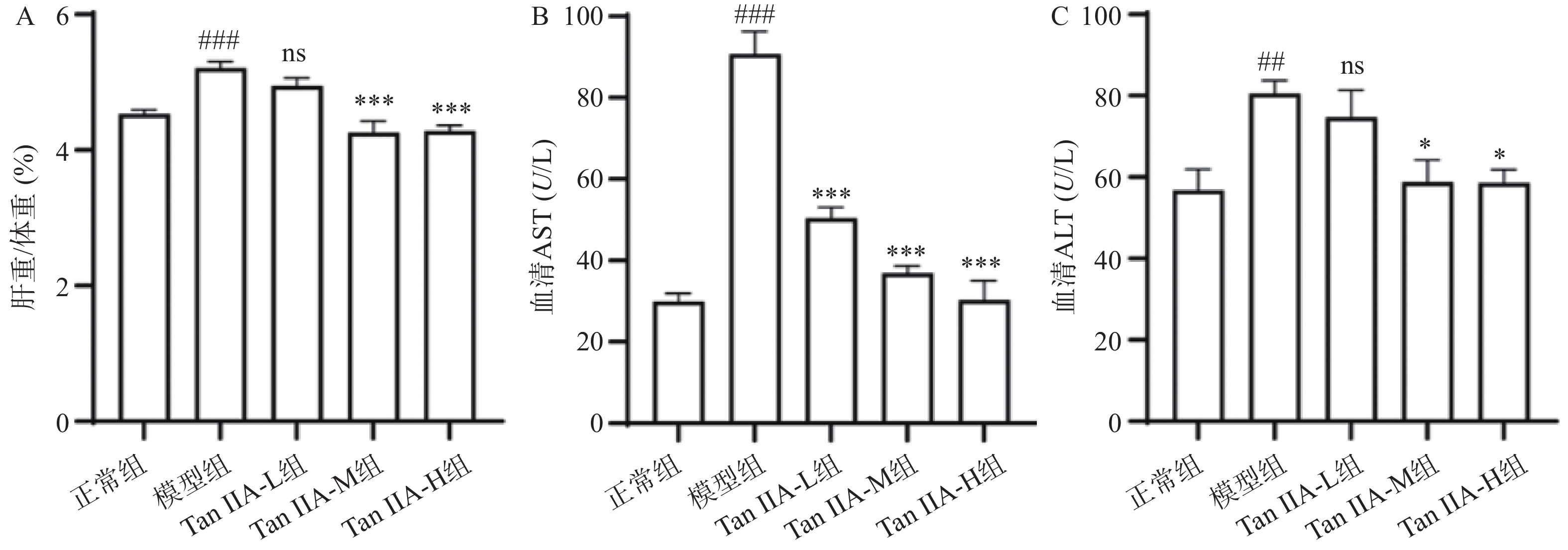
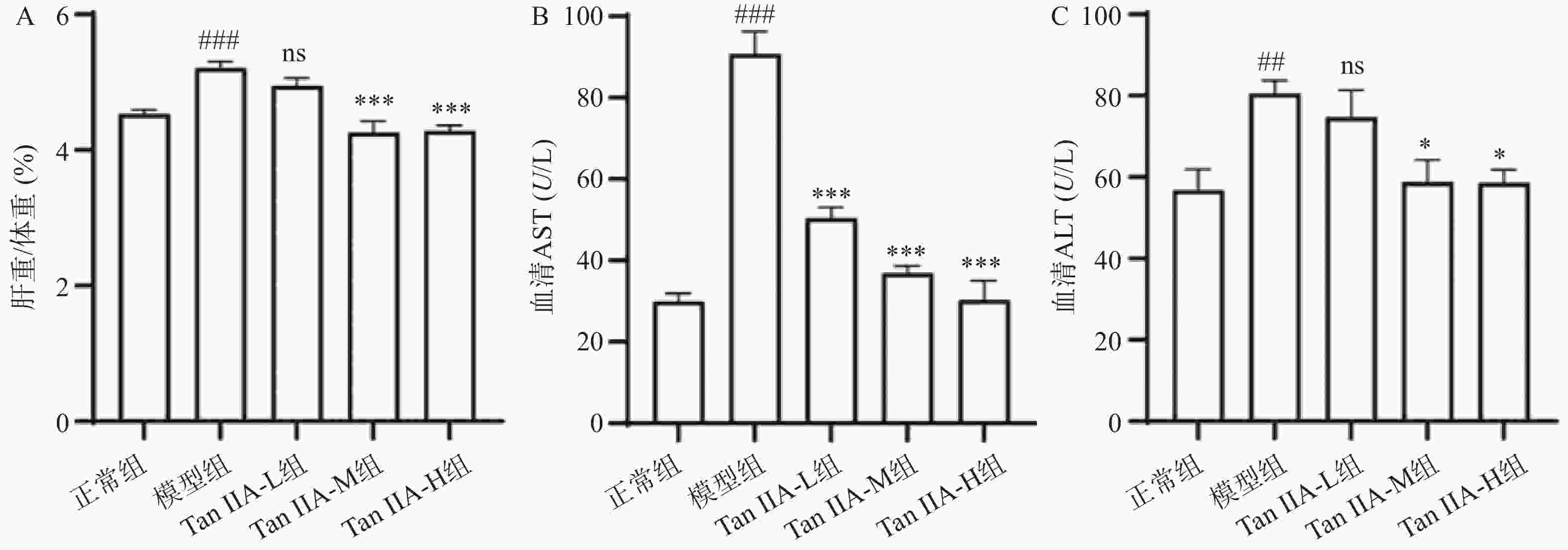
    • 与正常组相比,模型组小鼠的肝指数显著增加(P<0.001)。而给予丹参酮ⅡA后,只有中剂量组和高剂量组肝指数显著降低(P<0.001)。此外,APAP显着增加了血清ALT、AST水平(P<0.001),而中剂量及高剂量丹参酮ⅡA组预处理显著抑制了其升高(P<0.05)。这一结果清晰地表明,丹参酮ⅡA对于改善由肝损伤引起的血清ALT、AST水平升高具有显著效果。

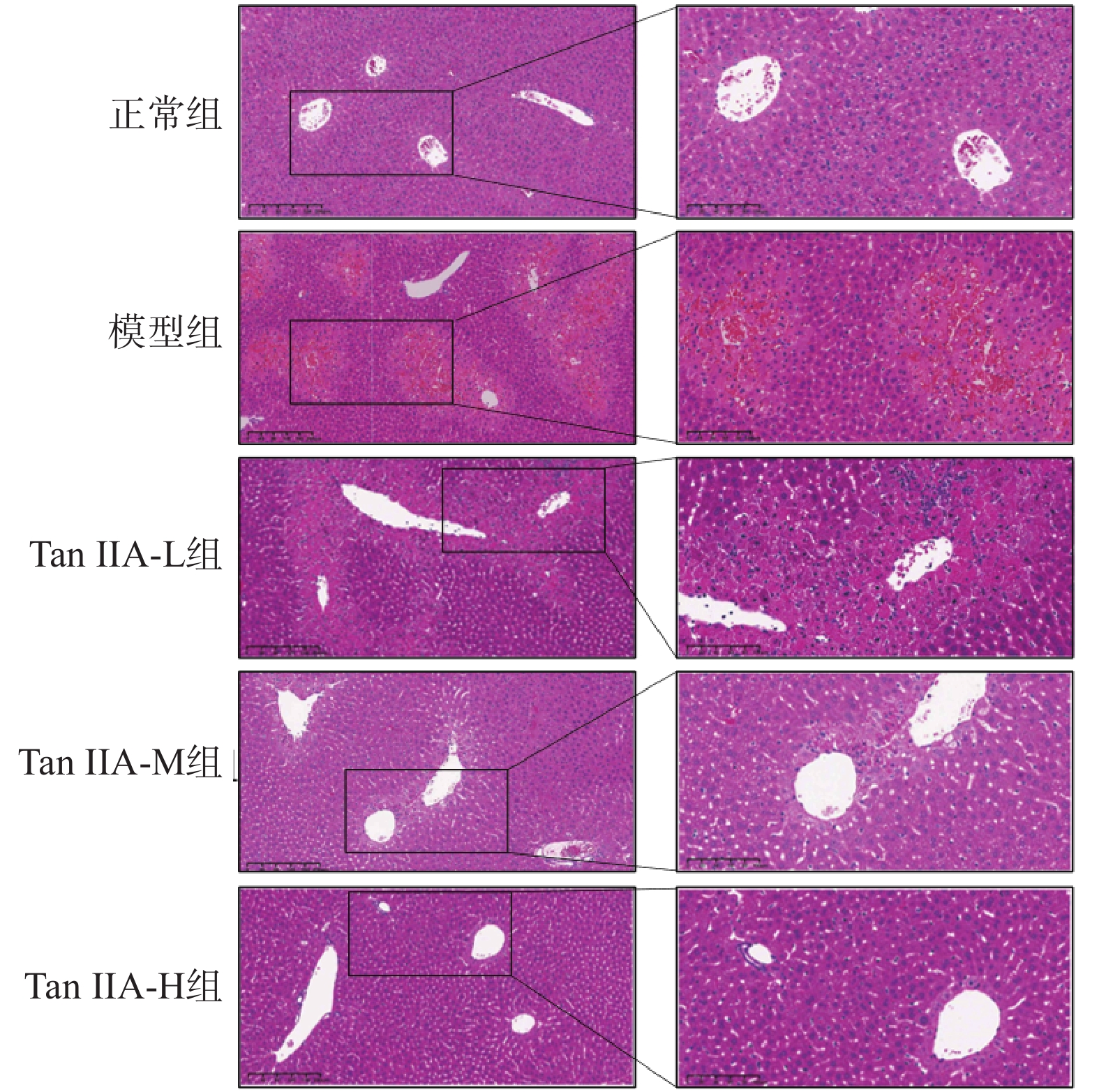
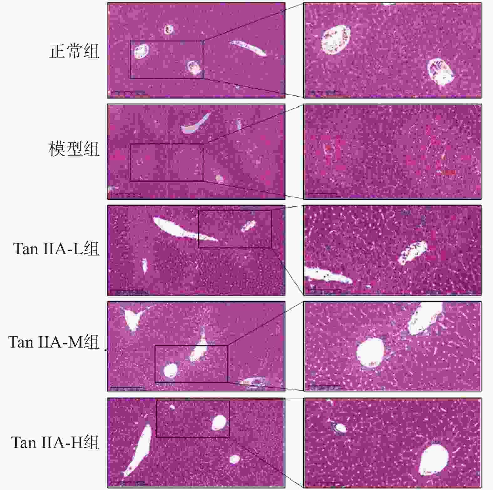
    • 在对照组小鼠的肝脏病理切片中(图2),可观察到肝细胞呈现出健康的形态学特征:肝细胞排列规则,肝小叶结构完整。而模型组小鼠的肝脏切片则出现明显病理变化:中央静脉周围的汇管区有大量的炎症细胞浸润,部分肝细胞的排列变得杂乱无章,并可见点状坏死区域。然而,在给予丹参酮ⅡA后,病理变化得到了显著的改善。具体表现为,低剂量丹参酮ⅡA组相较于模型组,肝脏细胞的炎症浸润程度有所减轻,显示出治疗带来的初步积极效应。相比之下,中剂量组与高剂量组的肝脏细胞排列恢复至正常状态,炎症细胞浸润也得到了明显的改善。这些观察结果共同为丹参酮ⅡA在肝损伤保护方面的潜在作用提供了有力的形态学证据。

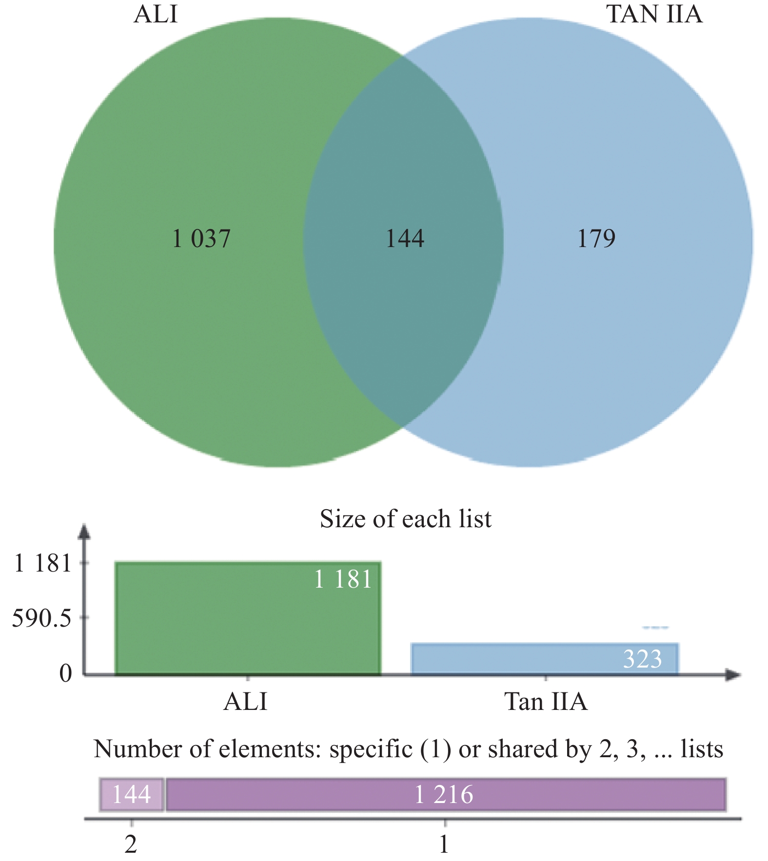
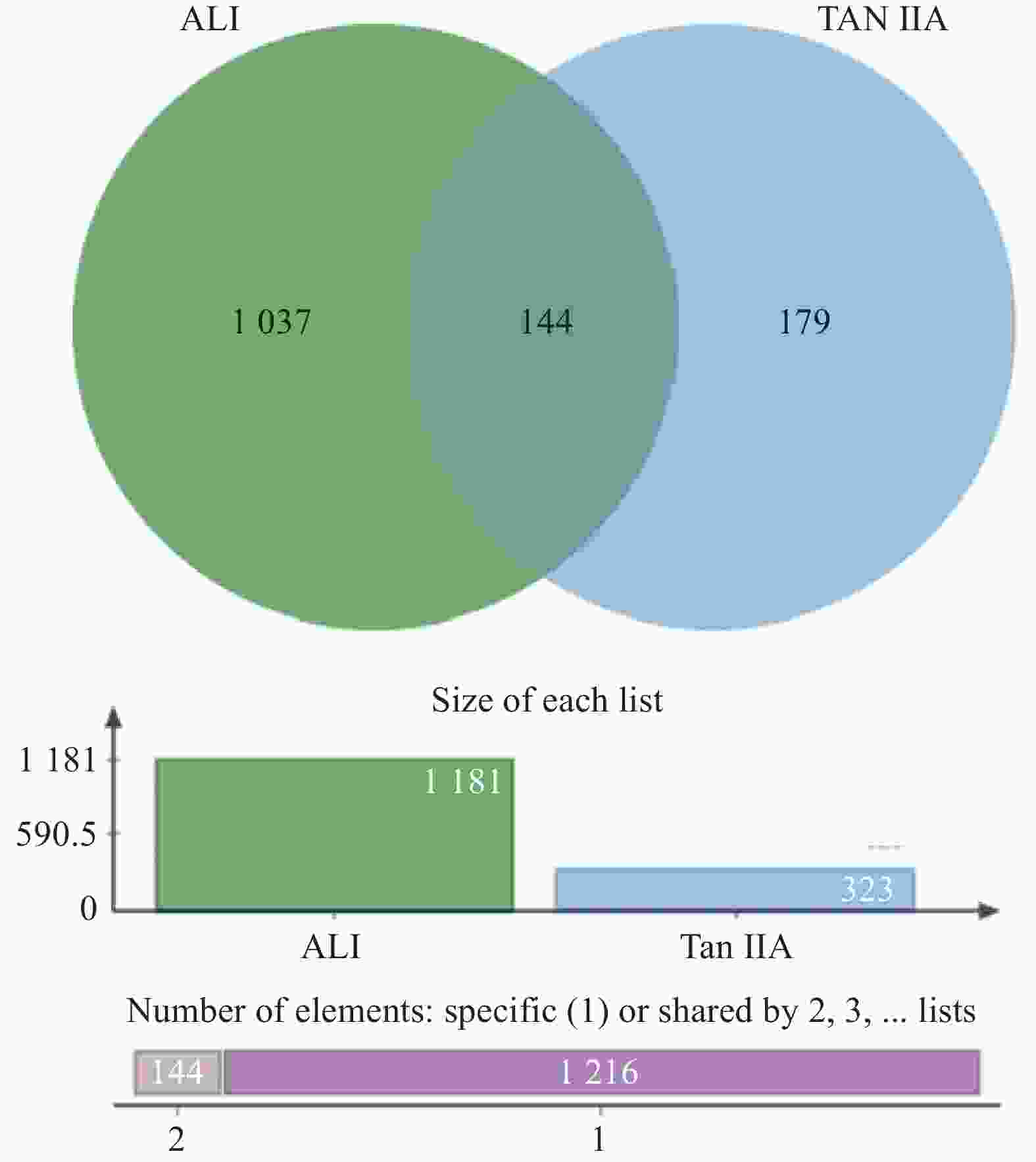
    • 根据ALI与丹参酮ⅡA的靶点韦恩图(图3)所示,共预测了1,181个ALI相关靶点,323个丹参酮ⅡA的作用靶点,交集靶点共144个。

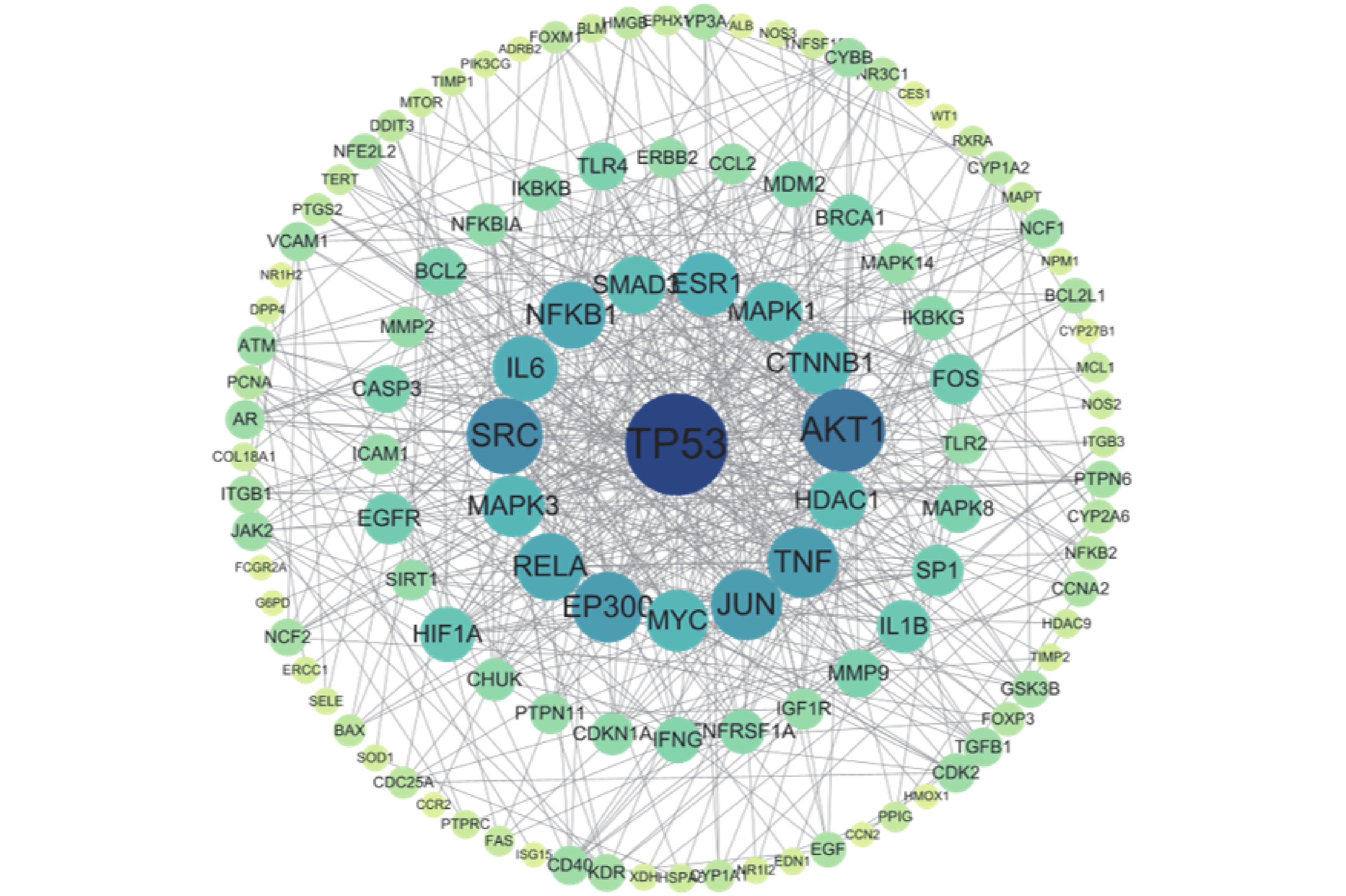
    • STRING数据库中分析显示,丹参酮ⅡA与ALI共同靶点的PPI图(图4)共得到545条边,PPI网络富集的P值小于1.0E−16。利用“Closeness unDir”、“Betweenness unDir”、“Degree unDir”Threshold值“0.0030726498027232726”、“228.3965517241378”、“9.362068965517242”筛选出关键靶点基因20个(见表1)。结果表明,TP53、AKT1、SRC、TNF和JUN可能是丹参酮ⅡA调控ALI的潜在靶点。
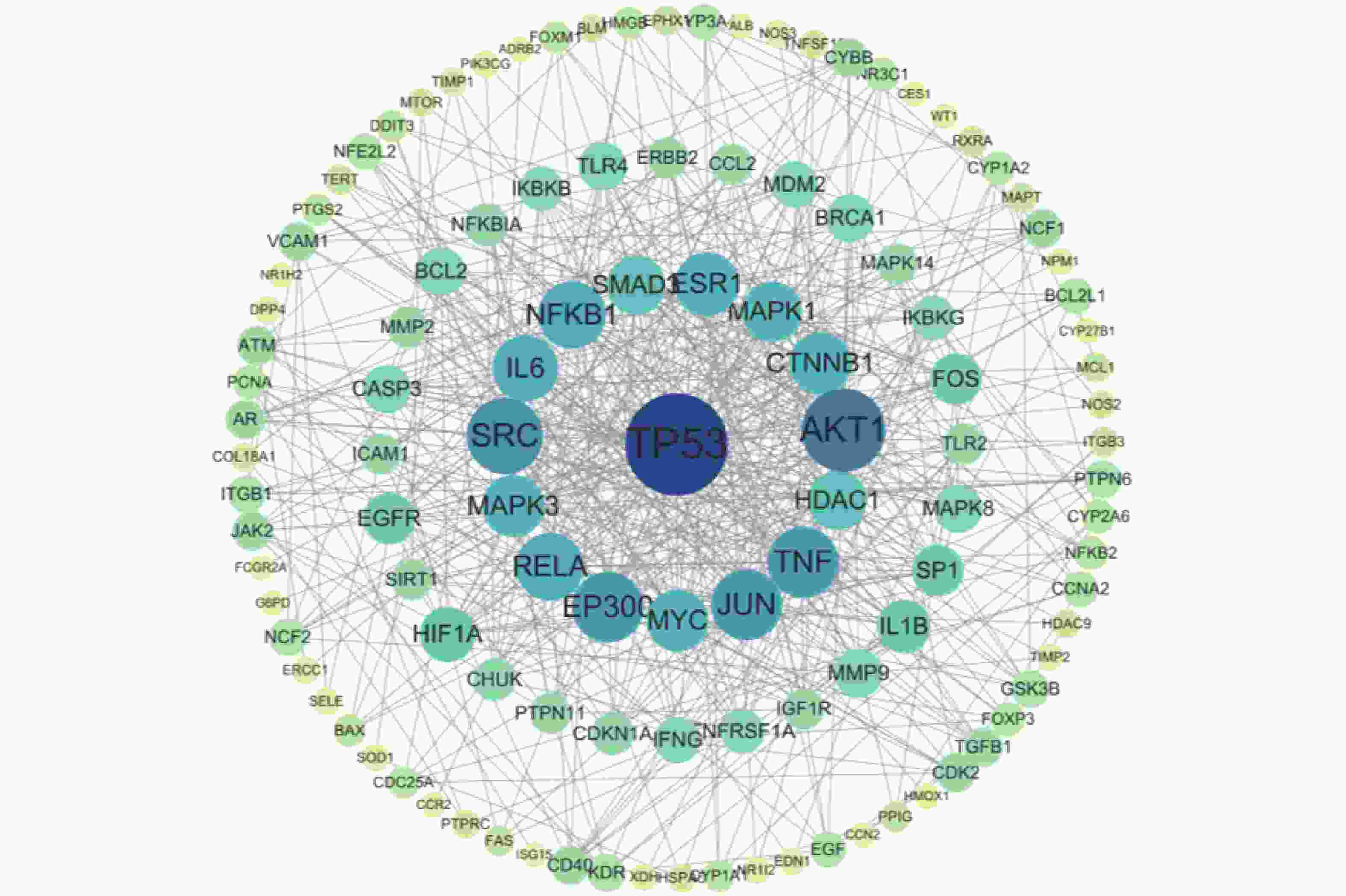
      序号基因名Betweenness unDirCloseness unDirDegree unDir
      1TP532 237.4860.004 3145
      2AKT11 760.5890.004 27434
      3SRC1 942.9690.004 09830
      4TNF894.444 30.003 86127
      5JUN595.766 40.004 18427
      6EP3003 048.2990.004 27427
      7RELA500.184 70.003 86125
      8NFKB1740.388 30.003 92225
      9IL6728.118 80.003 6124
      10CTNNB1443.343 70.003 98422
      11MYC284.3740.003 83122
      12MAPK3337.700 70.003 84622
      13MAPK1291.691 80.003 80221
      14SMAD3274.065 20.003 78820
      15EGFR326.358 60.003 6116
      16MMP9484.543 20.003 23614
      17BRCA1269.079 20.003 44814
      18BCL2281.804 90.003 3914
      19CASP3230.748 60.003 59714
      20CCL2253.517 70.003 26810
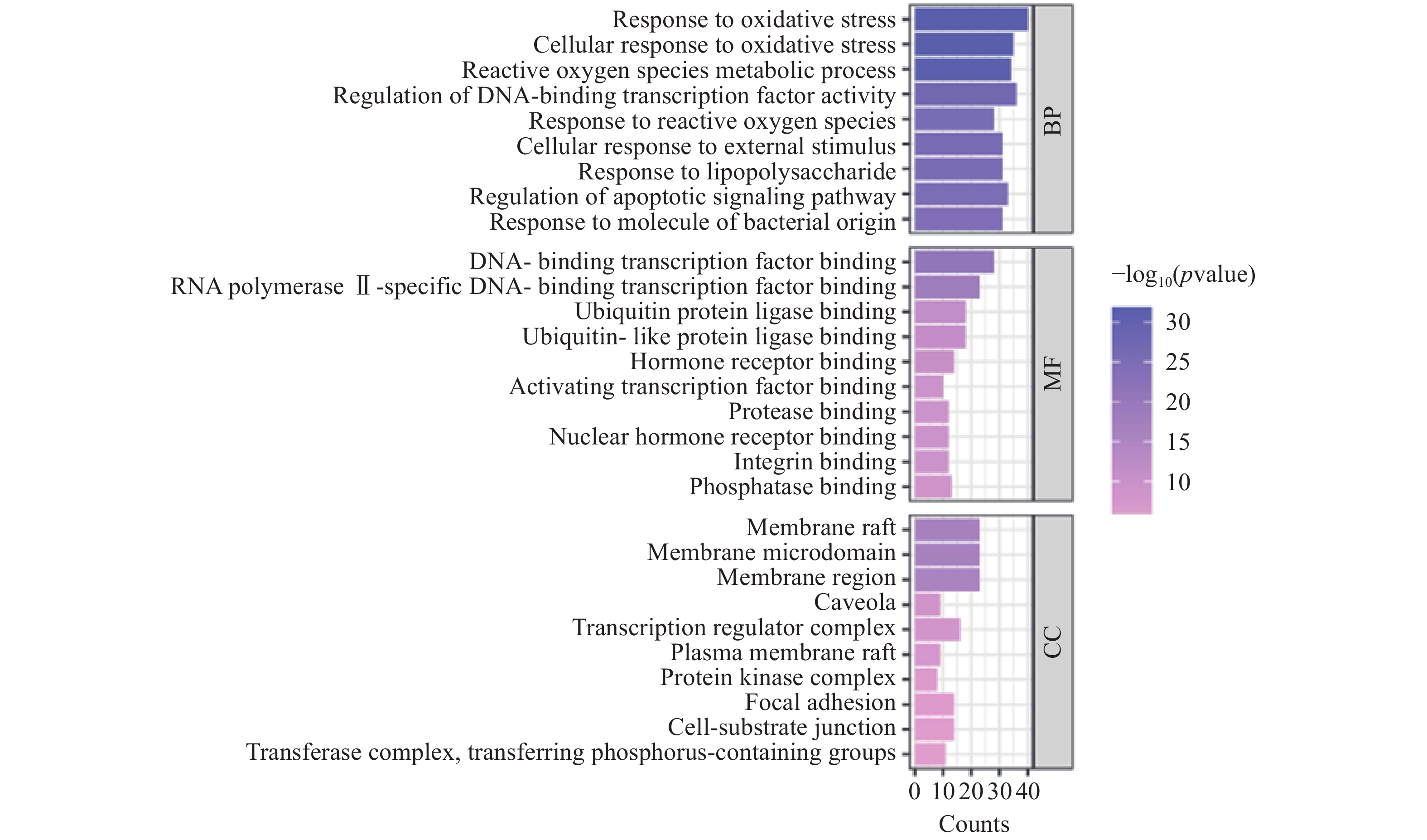
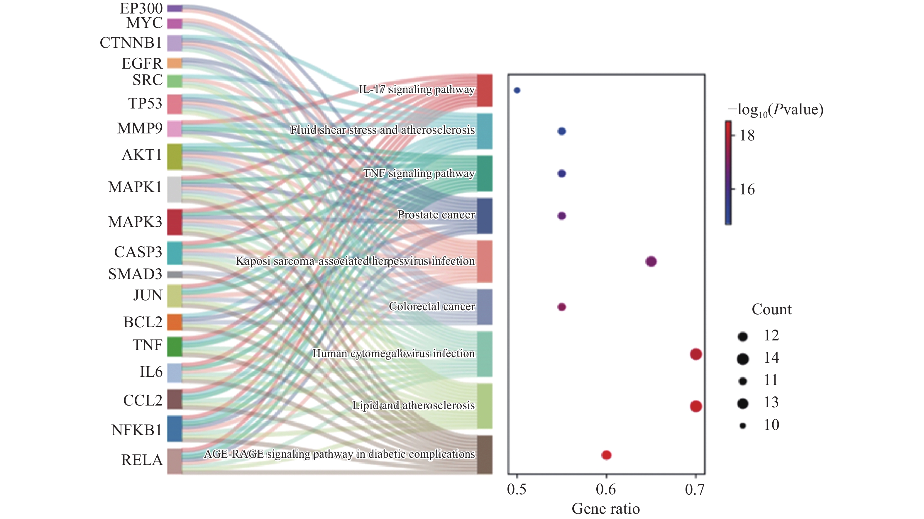
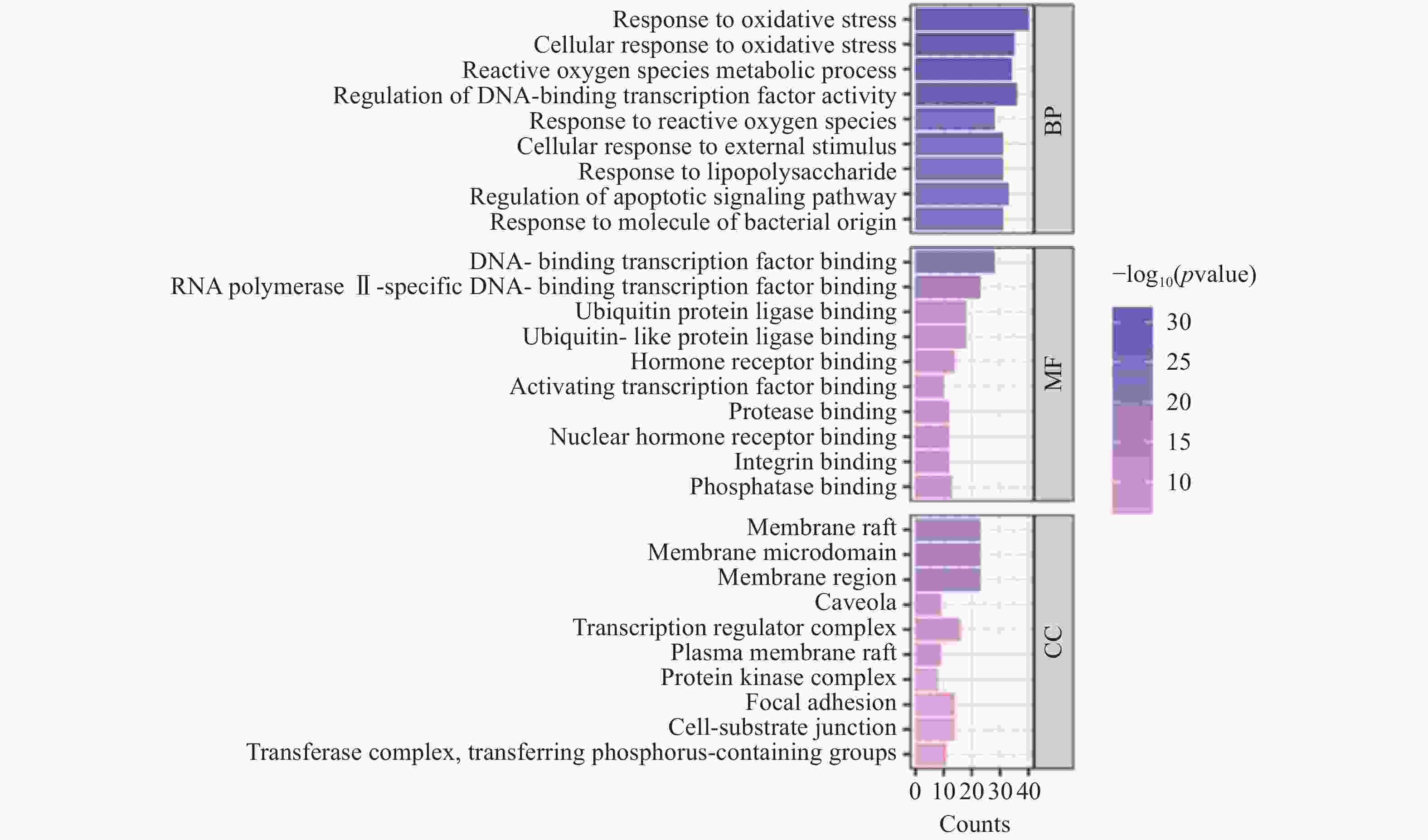
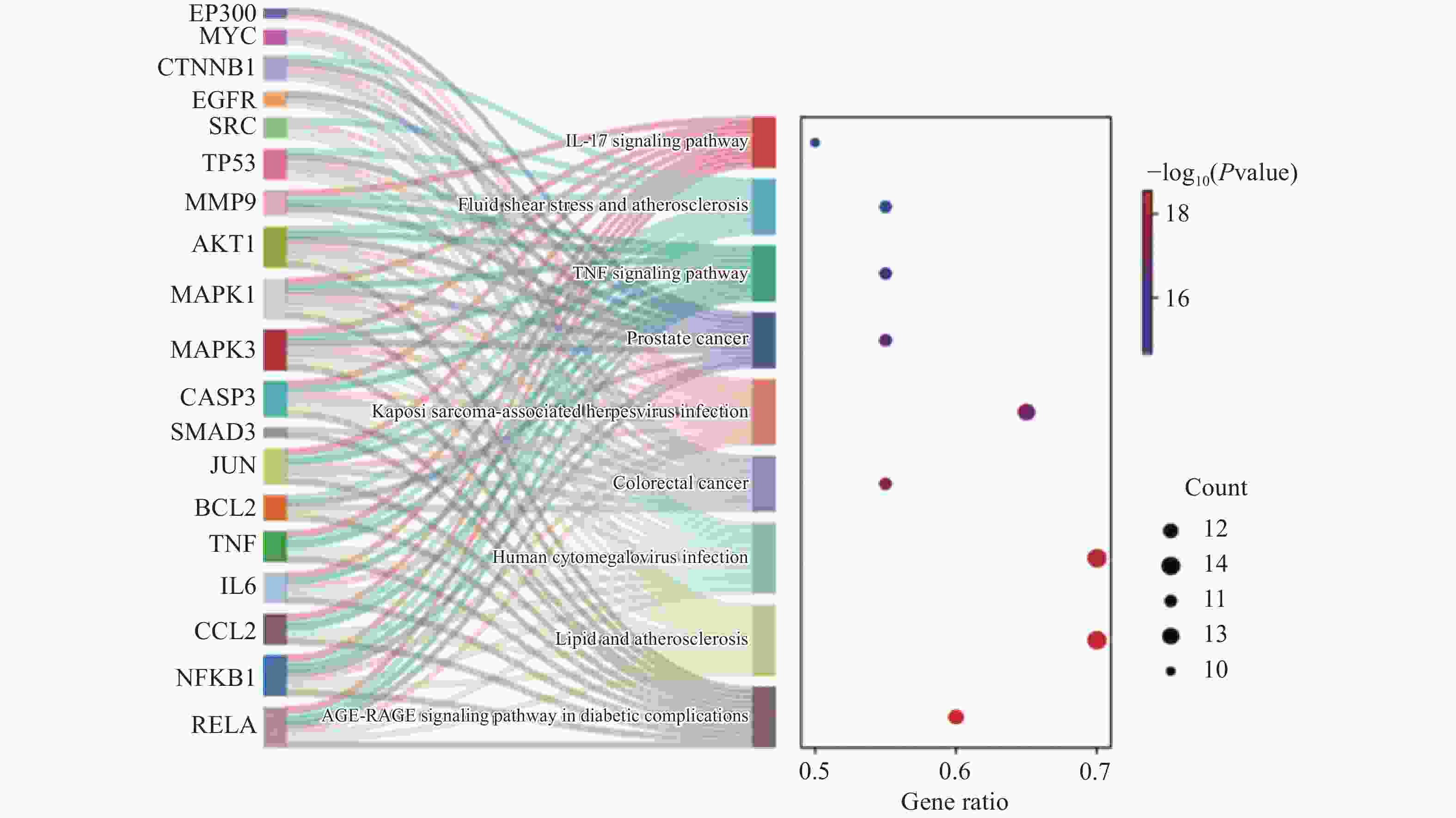
    • 为了更深入地探究丹参酮ⅡA改善ALI的作用机制,我们利用微生信平台对20个核心靶点进行了GO和KEGG富集分析,并生成了相应的条形图及桑葚图以直观展示结果。

      图5的GO富集分析显示,丹参酮ⅡA调控ALI主要涉及氧化应激反应、活性氧代谢过程的调控以及DNA结合转录因子活性的调节紧密相关等生物过程(BP);DNA转录因子的结合能力、泛素蛋白连接酶的结合活性等分子功能(MF);细胞膜的构成与结构特征等细胞成分(CC)。如图6所示,我们还筛选了与核心靶点相关的信号通路,主要涉及9条KEGG通路,包括IL-17信号通路、TNF信号通路、糖尿病并发症中的AGE-RAGE信号通路等,。

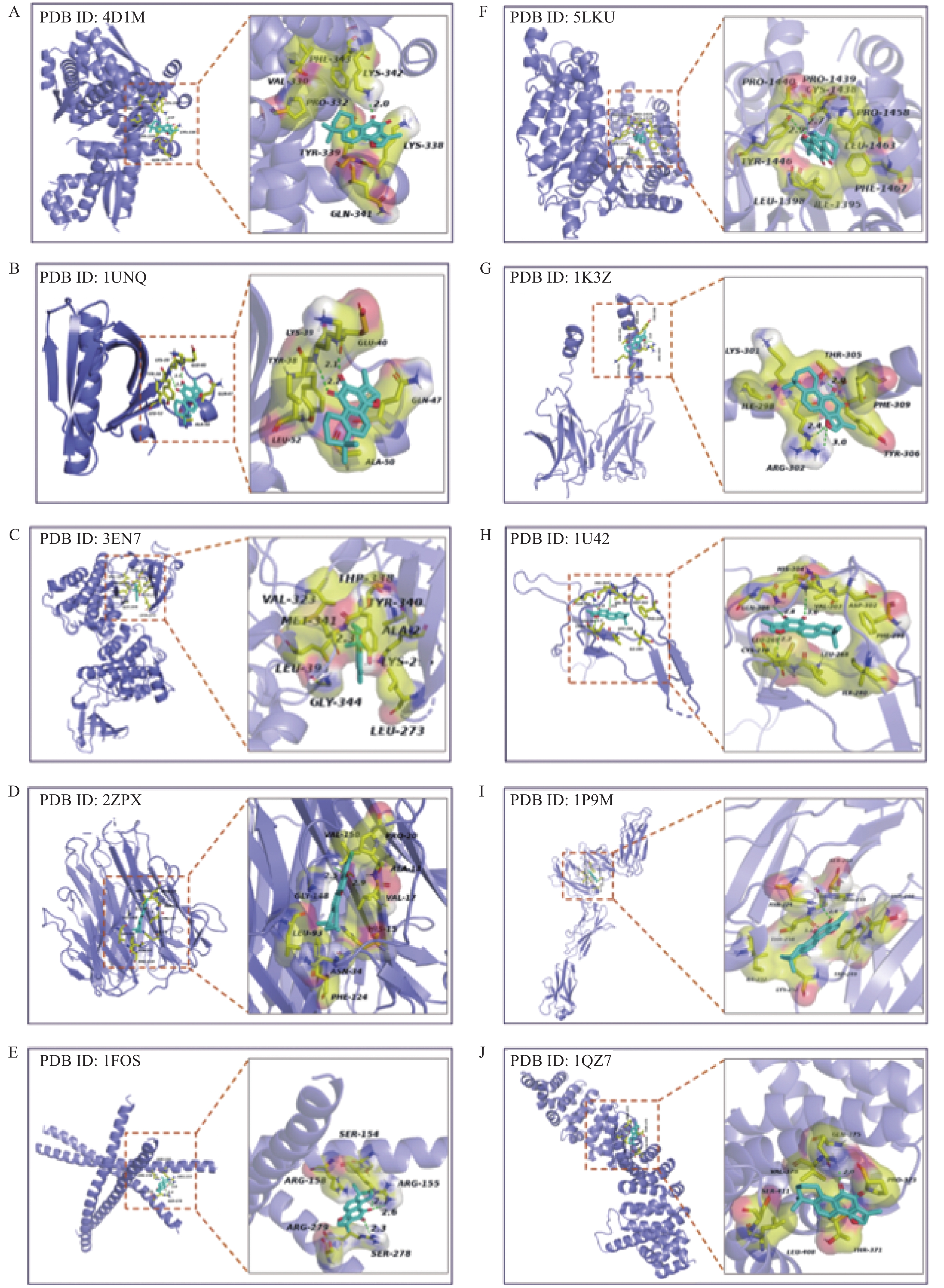
    • 在研究中,我们筛选出20个重要的核心靶点作为丹参酮ⅡA对ALI潜在治疗作用的关键参与者。为了进一步研究丹参酮ⅡA对ALI的治疗作用机制,我们利用分子对接模拟工具Autodock评估了丹参酮ⅡA与其可能的靶蛋白之间的结合相互作用(表2)。分子模拟结果表明,丹参酮ⅡA与MMP9、NFKB1、TNF、EP300和SMAD3等核心靶点之间具有较强的结合亲和力(图7)。此外,丹参酮ⅡA能与参与调控ALI的关键靶点TP53、AKT1、SRC、TNF和JUN等结合。表明丹参酮ⅡA可能与TP53、AKT1、SRC、TNF和JUN等靶点相互作用调控ALI。

      关键靶点PDB ID对接分数(kcal/mol)关键靶点PDB ID对接分数(kcal/mol)
      TP534D1M−5.74MYC5I4Z−6.64
      AKT11UNQ−6.71MAPK32ZOQ−7.29
      SRC3EN7−7.29MAPK11WZY−6.05
      TNF2ZPX−8.82SMAD31MK2−7.39
      JUN1FOS−5.25EGFR1M17−6.21
      EP3005LKU−7.85MMP91L6J−9.92
      RELA1K3Z−6.56BRCA11L0B−7.17
      NFKB11U42−8.06BCL22W3L−6.67
      IL61P9M−6.70CASP31NMQ−7.06
      CTNNB11QZ7−5.42CCL23IFD−6.65
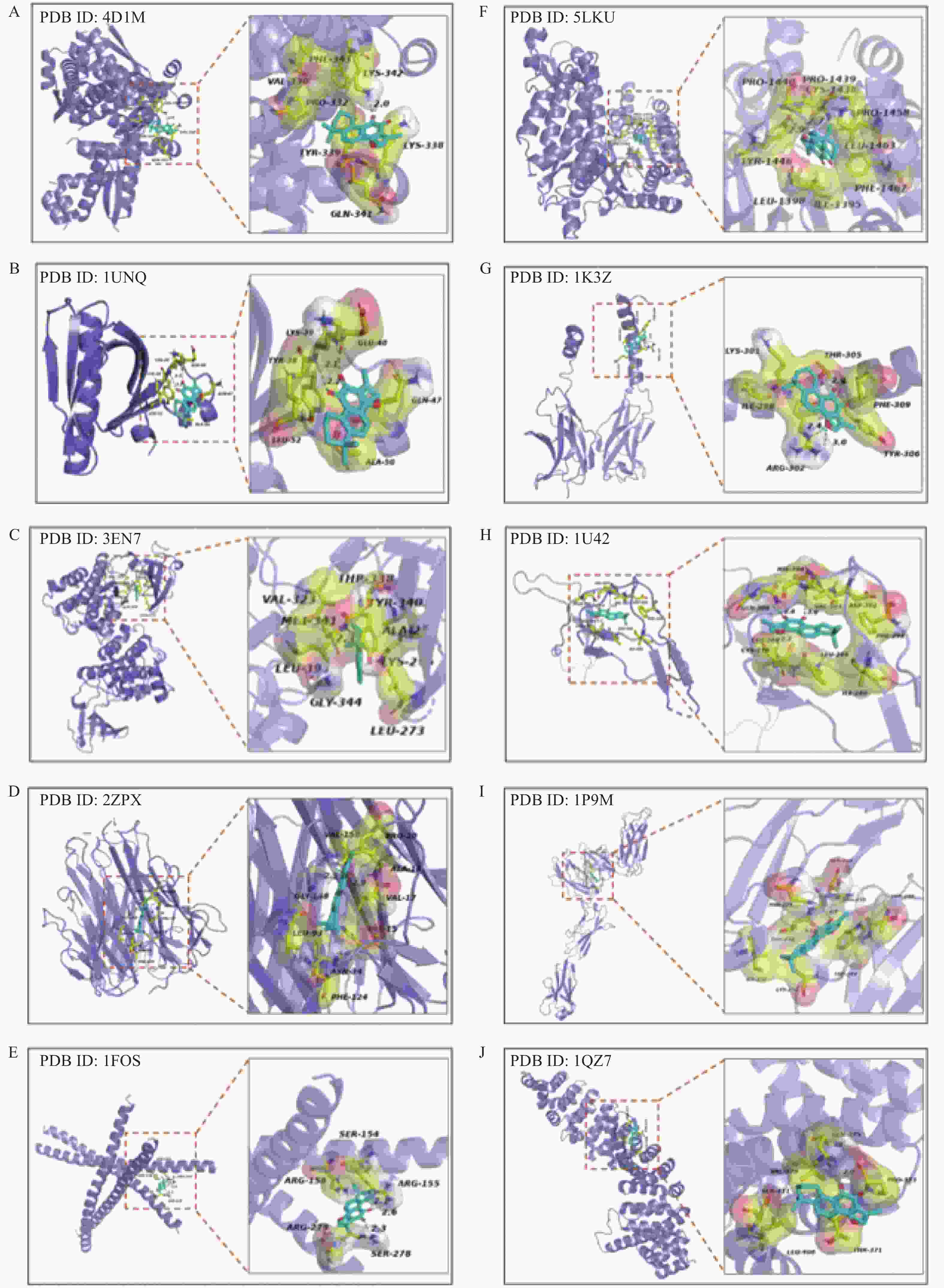
    • 肝损伤是世界公认的严重健康问题,现阶段临床治疗肝损伤的安全有效药物很少[2]。尽管已有研究指出丹参酮ⅡA对ALI具有改善作用[13-16],但其对APAP诱导DILI的治疗潜力及作用机制仍需进一步探究。本研究结合动物实验的肝指数、血清转氨酶水平及肝脏病理切片结果,验证了丹参酮ⅡA的保肝作用。我们发现模型组AST、ALT水平显著升高并且肝脏切片出现明显的病理变化,而在丹参酮ⅡA中、高剂量组,小鼠血清中的ALT和AST水平显著降低。此外,病理切片分析显示,丹参酮ⅡA中、高剂量组小鼠肝脏的空泡变性减少,细胞排列趋于正常,炎症细胞浸润减轻,进一步证实了丹参酮ⅡA对ALI小鼠肝脏结构的修复作用。低剂量组虽仅AST显著改善,且病理切片显示炎症有所减轻,表明肝脏正处于初步恢复阶段,这可能与给药时间短、剂量较低有关,导致其他生化指标尚未出现明显变化。

      此外,我们通过网络药理学分析发现了丹参酮ⅡA调控ALI的20个潜在靶点。PPI网络得出TP53、AKT1、SRC、TNF、JUN等靶点可能发挥着重要调控作用。将丹参酮ⅡA与部分靶点进行分子对接,发现丹参酮ⅡA与TP53、AKT1、SRC、TNF和JUN等靶点可能存在较强相互作用。肿瘤抑制基因TP53编码的p53蛋白在细胞周期的调控、细胞凋亡和衰老等多个重要生物学功能中发挥着关键作用[17, 18]。有研究表明,丹参酮ⅡA能够激活P53导致细胞凋亡和细胞周期停滞在G1/G0检查点,最终使HepG2细胞死亡[19]。磷脂酰肌醇3-激酶/蛋白激酶B(PI3K/AKT)信号通路通过对IκBα进行磷酸化和促进其降解,从而调节NF-κB的转录活性,该过程进一步介导炎症因子的生成和肝细胞的凋亡[20, 21]。丹参酮ⅡA能够通过PI3K/Akt/mTOR介导的抗炎、抗铁死亡和抗细胞凋亡来防止I/R诱导的急性肺损伤[22]。作为SFKs家族的一员,SRC与炎症和纤维化过程密切相关23, 24]。肿瘤坏死因子α(TNF-α)通过与其受体结合后激活caspase 3,从而导致肝细胞的凋亡[25]。过度氧化应激和c-Jun激活激酶的长期激活而导致肝功能异常[26]。其他靶点如Bcl2、CASP3、NF-κB在细胞凋亡、炎症反应等过程中发挥重要作用。

      KEGG通路富集分析结果显示,丹参酮ⅡA可能通过多条信号通路发挥保护肝脏的作用,包括IL-17信号通路、TNF信号通路以及糖尿病并发症中的AGE-RAGE信号通路。具体而言,白细胞介素17(IL-17)是一种促炎细胞因子,主要在招募炎症细胞,特别是中性粒细胞到炎症部位方面发挥关键作用[27-29]。相关研究指出,IL-17在APAP诱导的肝毒性中通过调节炎症反应扮演重要角色,这也意味着对IL-17进行抑制可能成为处理APAP诱导肝毒性的一个有效治疗策略[30]。有研究在小鼠银屑病模型中证实了丹参酮IIA具有免疫调节特性,并通过IL-17/IL-23和PTGS2/NF-κB/AP-1信号通路提供炎症保护[31]。同时,TNF-α也能激活NF-κB信号通路,引发IL-1β或IL-6等炎症因子的释放[32],这进一步加剧了肝脏的炎症反应[33]。丹参酮ⅡA已被证明能够通过下调NF-κB信号通路,抑制炎症因子的产生,从而防止脓毒症引起的肺损伤[34]。AGE与RAGE的结合会引发细胞内氧化应激,进而导致转录因子如NF-κB的激活[35],这会增强炎症反应并造成局部组织损伤[36]

      总而言之,丹参酮ⅡA可能通过与核心靶点TP53、AKT1、SRC、NF-kB和TNF的相互作用,调控TNF、IL-17等信号通路,从而改善ALI。本研究通过体内模型验证了丹参酮ⅡA对ALI的保护作用,并通过网络药理学及分子对接技术分析了其潜在的作用靶点及信号通路,揭示了丹参酮IIA保护肝脏的分子机制,为抗ALI药物研发提供了新思路。

Reference (36)

Catalog

    /

    DownLoad:  Full-Size Img  PowerPoint
    Return
    Return