Message Board

Respected readers, authors and reviewers, you can add comments to this page on any questions about the contribution, review,        editing and publication of this journal. We will give you an answer as soon as possible. Thank you for your support!

Name
E-mail
Phone
Title
Content
Verification Code
Volume 42 Issue 6
Jun.  2024
Turn off MathJax
Article Contents

YANG Yuanyuan, AN Xiaoqiang, XU Jiajie, JIANG Jian, LIANG Yuanyuan. Synergistic effect of positive electret combined with 5-fluorouracil on growth inhibition of scar fibroblasts[J]. Journal of Pharmaceutical Practice and Service, 2024, 42(6): 244-247. doi: 10.12206/j.issn.2097-2024.202310027
Citation: YANG Yuanyuan, AN Xiaoqiang, XU Jiajie, JIANG Jian, LIANG Yuanyuan. Synergistic effect of positive electret combined with 5-fluorouracil on growth inhibition of scar fibroblasts[J]. Journal of Pharmaceutical Practice and Service, 2024, 42(6): 244-247. doi: 10.12206/j.issn.2097-2024.202310027

Synergistic effect of positive electret combined with 5-fluorouracil on growth inhibition of scar fibroblasts

doi: 10.12206/j.issn.2097-2024.202310027
  • Received Date: 2023-10-16
  • Accepted Date: 2024-04-11
  • Rev Recd Date: 2024-03-19
  • Available Online: 2024-06-24
  • Publish Date: 2024-06-25
  •   Objective   To investigate the effects and possible mechanism of electret and 5-fluorouracil(5-FU)on the growth of scar fibroblasts.   Methods   The effect of +5000 V electret combined with different concentrations of 5-FU on the proliferation of scar fibroblasts was detected by automatic enzyme labeling instrument. The apoptosis of scar fibroblasts and the mRNA expression of p53 and other apoptotic genes were studied by fluorescence microscopy and RT-PCR technology under the action of electrostatic field.   Results   ① After the treatment of positive electret and different concentrations of 5-FU for 72 h, the cell proliferation rate decreased, and the inhibition rate of scar cells in the +5000 V electret+160 μg/ml 5-FU group was (0.15±0.051)%. ②+5000 V electret group could promote the apoptosis of scar fibroblasts; The number of apoptotic cells in +5000 V electret and 5-FU group was higher than that in 5-FU group. ③The mRNA expression levels of four apoptotic genes in the +5000 V electret group were increased, and the expression levels of four signature genes in the +5000 V electret and 5-FU group were increased compared with those in the 5-FU group.   Conclusion   The combination of positive electret and 5-FU had a synergistic effect on inhibiting cell growth. The mechanism of positive electret inhibiting scar cell growth may be through promoting the expression of apoptosis gene, and then affecting the growth state of cells to inhibit cell growth.
  • [1] 柳承业, 徐凯, 林瀚. 曲安奈德与A型肉毒毒素联合注射治疗瘢痕疙瘩的疗效观察[J]. 中国美容整形外科杂志, 2020, 31(7):430-432,453. doi:  10.3969/j.issn.1673-7040.2020.07.015
    [2] 安晓强, 苑旺, 梁媛媛, 等. 正极性驻极体5-氟尿嘧啶贴剂对兔耳瘢痕组织中Ⅰ、Ⅲ型胶原和TCF-β表达的影响[J]. 药学实践杂志, 2019, 37(2):115-120,145.
    [3] YAO G, MO X Y, YIN C H, et al. A programmable and skin temperature-activated electromechanical synergistic dressing for effective wound healing[J]. Sci Adv, 2022, 8(4):eabl8379. doi:  10.1126/sciadv.abl8379
    [4] LIANG Y Y, XU J J, SUN Z P, et al. Research progress on the correlation between bacterial biofilm microenvironment and charge regulation[J]. IEEE Trans Dielectr Electr Insul, 2022, 29(4):1540-1545. doi:  10.1109/TDEI.2022.3183663
    [5] LUO R Z, SHI B J, LUO D, et al. Self-powered electrical stimulation assisted skin wound therapy[J]. Sci Bull, 2023, 68(16):1740-1743. doi:  10.1016/j.scib.2023.07.017
    [6] VERDES M, MACE K, MARGETTS L, et al. Status and challenges of electrical stimulation use in chronic wound healing[J]. Curr Opin Biotechnol, 2022, 75:102710. doi:  10.1016/j.copbio.2022.102710
    [7] LI X, WANG Y R, XU M X, et al. Polymer electrets and their applications[J]. Appl Polym Sci, 2021, 20: 50406.
    [8] SUN Z P, WANG H B, GUO X, et al. Electret prevents the formation of bacterial biofilm[J]. IET Nanodielectr, 2023, 6(2):57-63. doi:  10.1049/nde2.12051
    [9] 梁媛媛, 涂晔, 崔黎丽, 等. 驻极体静电场对巨噬细胞迁移能力的影响[J]. 第二军医大学学报, 2021, 42(6):688-692.
    [10] STRATFORD J P, EDWARDS C L A, GHANSHYAM M J, et al. Electrically induced bacterial membrane-potential dynamics correspond to cellular proliferation capacity[J]. Proc Natl Acad Sci U S A, 2019, 116(19):9552-9557. doi:  10.1073/pnas.1901788116
    [11] 王碧瑶, 肖含先之, 牛依琳, 等. 当归多糖促进5-氟尿嘧啶作用后小鼠应激性红细胞发生[J]. 中国药理学通报, 2023, 39(10):1949-1956. doi:  10.12360/CPB202211087
    [12] 田阳, 王飞, 徐骁然, 等. 沉默单核细胞趋化蛋白3对人增生性瘢痕成纤维细胞增殖、迁移及凋亡的影响[J]. 临床皮肤科杂志, 2022, 51(9):518-523.
    [13] ZHAO M. Electrical fields in wound healing-An overriding signal that directs cell migration[J]. Semin Cell Dev Biol, 2009, 20(6):674-682. doi:  10.1016/j.semcdb.2008.12.009
    [14] LUO R Z, DAI J Y, ZHANG J P, et al. Accelerated skin wound healing by electrical stimulation[J]. Adv Healthc Mater, 2021, 10(16):e2100557. doi:  10.1002/adhm.202100557
    [15] NUCCITELLI R. A role for endogenous electric fields in wound healing[J]. Curr Top Dev Biol, 2003, 58:1-26.
  • 加载中
通讯作者: 陈斌, bchen63@163.com
  • 1. 

    沈阳化工大学材料科学与工程学院 沈阳 110142

  1. 本站搜索
  2. 百度学术搜索
  3. 万方数据库搜索
  4. CNKI搜索

Figures(1)  / Tables(3)

Article Metrics

Article views(659) PDF downloads(1) Cited by()

Related
Proportional views

Synergistic effect of positive electret combined with 5-fluorouracil on growth inhibition of scar fibroblasts

doi: 10.12206/j.issn.2097-2024.202310027

Abstract:   Objective   To investigate the effects and possible mechanism of electret and 5-fluorouracil(5-FU)on the growth of scar fibroblasts.   Methods   The effect of +5000 V electret combined with different concentrations of 5-FU on the proliferation of scar fibroblasts was detected by automatic enzyme labeling instrument. The apoptosis of scar fibroblasts and the mRNA expression of p53 and other apoptotic genes were studied by fluorescence microscopy and RT-PCR technology under the action of electrostatic field.   Results   ① After the treatment of positive electret and different concentrations of 5-FU for 72 h, the cell proliferation rate decreased, and the inhibition rate of scar cells in the +5000 V electret+160 μg/ml 5-FU group was (0.15±0.051)%. ②+5000 V electret group could promote the apoptosis of scar fibroblasts; The number of apoptotic cells in +5000 V electret and 5-FU group was higher than that in 5-FU group. ③The mRNA expression levels of four apoptotic genes in the +5000 V electret group were increased, and the expression levels of four signature genes in the +5000 V electret and 5-FU group were increased compared with those in the 5-FU group.   Conclusion   The combination of positive electret and 5-FU had a synergistic effect on inhibiting cell growth. The mechanism of positive electret inhibiting scar cell growth may be through promoting the expression of apoptosis gene, and then affecting the growth state of cells to inhibit cell growth.

YANG Yuanyuan, AN Xiaoqiang, XU Jiajie, JIANG Jian, LIANG Yuanyuan. Synergistic effect of positive electret combined with 5-fluorouracil on growth inhibition of scar fibroblasts[J]. Journal of Pharmaceutical Practice and Service, 2024, 42(6): 244-247. doi: 10.12206/j.issn.2097-2024.202310027
Citation: YANG Yuanyuan, AN Xiaoqiang, XU Jiajie, JIANG Jian, LIANG Yuanyuan. Synergistic effect of positive electret combined with 5-fluorouracil on growth inhibition of scar fibroblasts[J]. Journal of Pharmaceutical Practice and Service, 2024, 42(6): 244-247. doi: 10.12206/j.issn.2097-2024.202310027
  • 损伤部位的机械刺激和局部感染可导致皮肤伤口的异常愈合,甚至导致瘢痕疙瘩的形成[1]。细胞的迁移、生长和增殖在伤口修复过程中起着至关重要的作用, 增生性瘢痕的形成与各类因子表达异常、成纤维细胞的增殖过度、以及胶原的过度沉积等因素密切相关[2-3]。电生理学实验研究表明,生物组织的行为与电荷的流动高度相关[4-6]

    驻极体是一类不需外加电源即能够提供稳定静电场和微电流的功能电介质材料[7], 其产生的静电场能够加快受创部位微循环,促进成纤维细胞在不同时期生长或凋亡以及促进药物的经皮渗透等作用[8-10]。 5-氟尿嘧啶(5-FU)是最早研发上市的抗癌药,作用于细胞后抑制其 DNA的合成,从而对成纤维细胞生长增殖产生影响,临床上是治疗难治性瘢痕的常用药之一[11]

    该研究将驻极体和 5-FU 联用,通过探索驻极体静电场联合5-FU对分离培养瘢痕成纤维细胞的生长、细胞周期和凋亡的影响,以期在细胞水平研究驻极体静电场以及驻极体联用 5-FU 对瘢痕形成的影响机制,为预防和治疗病理瘢痕提供新的思路。

    • Sprague-Dawley(SD)大鼠,雌雄不限,体质量为(180±20) g,购自海军军医大学实验动物中心,动物合格证号:SCXK(沪)2018-0006。

    • 水合氯醛(中国医药集团化学试剂有限公司);5-FU(上海生物工程有限公司);cck-8 试剂盒[东仁化学科技(上海)有限公司];Hoechst33342(上海如吉生物科技发展有限公司);引物(武汉擎科创新生物科技有限公司);细胞培养箱(Thermo公司);栅控恒压电晕充电系统( 复旦中学校办厂);ESR102A 型振动电容静电计(北京华晶汇科技有限公司);荧光定量 PCR 仪(ABI公司); 全自动定量酶标仪(Bio-Rad公司)。

    • 通过栅控恒压电晕充电系统对双裸面聚丙烯(Polypropylene,PP) 膜进行注极,栅压设为+5000 V,充电时间为 5 min,制备得+5000 V 驻极体。驻极体等效表面电位通过表面电位计(ESR102A 型振动电容静电计)测量。

    • 用 10%的水合氯醛腹腔注射麻醉(4 ml/kg),用实验动物剃毛刀去除大鼠背部毛发, 在大鼠背部用打孔器制造左右对称共 4 个直径为 2 cm 的创面,创面之间间隔一定距离,去除肉膜层,分笼饲养。在创面形成后 4 周产生增生性瘢痕。

    • 取瘢痕模型大鼠,10%的水合氯醛腹腔注射麻醉, 用实验动物剃毛刀将大鼠背部毛发去除干净, 酒精棉球擦拭后, 手术剪取下瘢痕皮肤组织,组织用含有青霉素浓度 100 U/ml,链霉素浓度 100 μg/ml 的 PBS 缓冲液反复冲洗 3 次。冲洗后, 用眼科剪将瘢痕皮肤组织剪成组织小块, 组织小块大小在 1~2 mm3 之间。将组织小块置于培养皿中,加入含有 20%胎牛血清的 1640培养基,待细胞长满培养皿,进行细胞传代,待细胞传至 3~8 代,进行后续实验。

    • 实验分为瘢痕细胞对照组,+5000 V驻极体组,+5000 V驻极体+10 μg/ml 5-FU组,+5000 V驻极体+40 μg/ml 5-FU组,+5000 V驻极体+160 μg/ml 5-FU组。

    • 取对数生长期的瘢痕皮肤组织成纤维细胞(细胞密度为 1×10⁴/ 孔),接种于96孔培养板中(100 μl/孔) 。待细胞绝大部分贴壁后,更换培养液,按照上述实验分组分别对细胞干预24 h、48 h、72 h,再以 10 μl/孔向各孔中加入 cck-8 试剂溶液, 继续在细胞培养箱中保温 1~4 h,终止培养,震荡摇匀,在全自动定量酶标仪上以 450 nm 波长处测定各孔吸光度, 按照增殖率=(实验孔实测值−空白组平均值)/(对照组平均值−空白组平均值)×100%计算细胞增殖率。

    • 取瘢痕细胞消化离心后重悬,制成单细胞悬液,按1×10⁴个/孔种植到96孔板中,待细胞绝大部分贴壁后,更换培养基,选取+5000 V驻极体+40 μg/ml 5-FU组对其进行处理,恒温箱培养48 h后,去除培养液,各组均加入含Hoechst 33342(原浓度为10 mg/ml,稀释10000倍)的1640培养液,避光孵育30 min,然后去除培养液,用PBS清洗两次,加入适量PBS,置于荧光显微镜下观察并拍照,每组重复3个样本。荧光定量PCR引物序列表见表1

      引物名称引物序列(5'→3')片段长度(bp)退火温度(℃)
      R-TP53-SGAAGCCCTCCAAGTGTCAGC22060
      R-TP53-AGGCAGAACAGCTTATTGAGGGA60
      R-fas-SAGCGTTCGTGAAACCGACAAC17260
      R-fas-AAGTGTTTCCTGTCCGTGTACTCC60
      R-fasl-SGCAAATAGCCAACCCCAGCAC18660
      R-fasl-AACGAAGTACAACCCAGCCTCA60
      R-BAX-SGGGCCTTTTTGCTACAGGGTTT28460
      R-BAX-AAGCAAAGTAGAAAAGGGCAACCAC60
      R-GAPDH-SCTGGAGAAACCTGCCAAGTATG13860
      R-GAPDH-AGGTGGAAGAATGGGAGTTGCT60
    • 瘢痕细胞在不同浓度5-FU及正极性驻极体和不同浓度5-FU联用干预 24 h、48 h、72 h后增殖率变化情况:在5-FU干预 24 h、48 h、72 h后,在不同浓度5-FU的作用下, 随着 5-FU浓度的增加,瘢痕细胞的增殖率呈下降趋势,且随着时间的增加瘢痕增值下降趋势越明显;正极性驻极体和不同浓度5-FU联用作用于细胞72 h后,细胞增殖率都有所降低。随着 5-FU 浓度的增加, 对照组和各正极性驻极体组瘢痕细胞增殖率呈下降趋势;该变化趋势较5-FU单一作用 72 h组更加明显,具体见表2

      组别01040160
      24 h组1.00±0.0280.99±0.0280.77±0.0270.49±0.033
      48 h组1.00±0.0240.98±0.0250.65±0.0280.41±0.028
      72 h组1.00±0.0200.96±0.0330.53±0.0170.21±0.046
      +5000 V与5-FU联用72 h组0.90±0.0340.47±0.0510.15±0.051
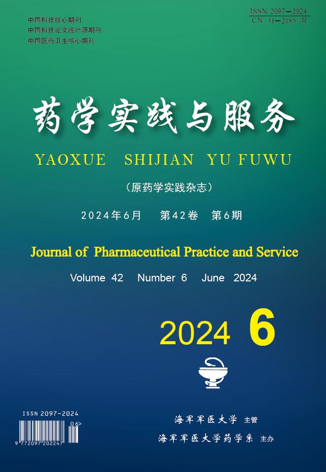
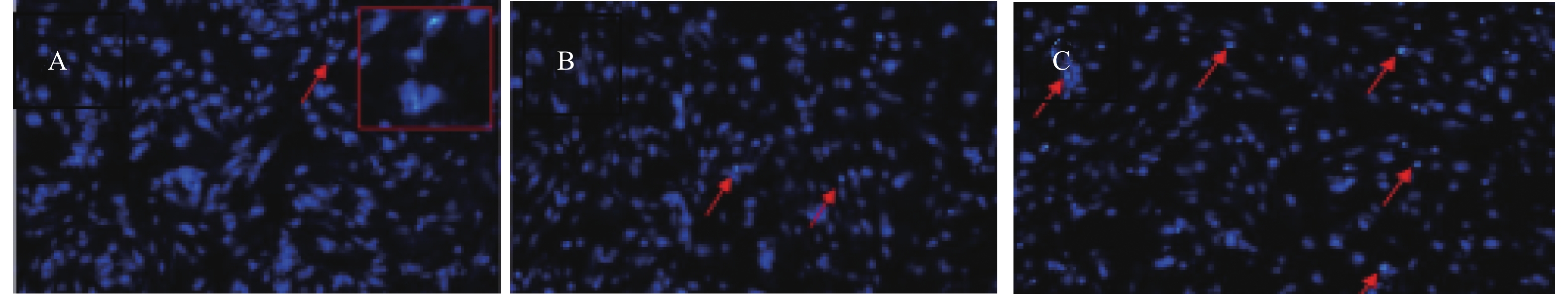
    • 瘢痕细胞各组染色的结果见图1,图中箭头所示亮点为凋亡细胞,较暗的蓝色荧光为正常细胞,图1A右上角方框内为亮点部位放大结果。结果显示:瘢痕细胞对照组基本无细胞凋亡或者极少细胞凋亡; +5000 V驻极体组出现少数细胞凋亡; +5000 V 驻极体与5-FU联用组细胞凋亡数高于 5-FU 组。

    • 瘢痕成纤维细胞各组凋亡基因 mRNA 的相对表达量见表3。以对照组为 1,比较各实验组mRNA 的表达。结果显示:与对照组相比,5-FU组表达量明显增加, +5000 V 驻极体组4种凋亡基因 mRNA 的相对表达量都有所增加;与5-FU组相比,虽然+5000 V驻极体组相对表达量稍小,但是+5000 V驻极体+5-FU组表达量则显著增大,以上相比较的各组之间差异均有统计学意义(P<0.05)。

      组别 p53 fas Bas fasl
      对照组 1.00±0.00 1.00±0.01 1.00±0.02 1.00±0.03
      5-FU组 1.60±0.06 2.23±0.11 1.81±0.13 2.01±0.19
      +5000 V 驻极体组 1.30±0.10 1.61±0.21 1.41±0.10 1.50±0.12
      +5000 V+5-FU组 1.92±0.14 2.81±0.29 2.20±0.17 2.49±0.21
    • 细胞的增殖和凋亡状况能够从细胞层面反应组织的生长状况[12]。实验结果表明:不同浓度的5-FU对瘢痕细胞的生长有不同的抑制作用,随着化学药物浓度的增加,瘢痕细胞的增值率降低更迅速。+5000 V驻极体与5-FU联用,对细胞的增殖有协同抑制作用,正极性驻极体和不同浓度5-FU联用作用于细胞72 h后,细胞增殖率均较5-FU单一作用于瘢痕细胞72 h的细胞增值率有所降低。且随着联合作用组5-FU浓度的增加,其抑制细胞增殖率作用更加明显,经+5000 V驻极体与160 μg/ml 5-FU联用72 h,对瘢痕细胞的抑制率可达0.15±0.051。

      细胞凋亡检测实验也表明,由于驻极体静电场的作用,使瘢痕细胞凋亡数量增加,+5000 V驻极体组凋亡细胞增多,+5000 V驻极体与5-FU联用组细胞凋亡数高于5-FU组。p53、Fas、Fasl、Bax 4种基因的表达结果也证明+5000 V 驻极体能够通过一定程度的影响该4种基因的表达来促进细胞凋亡,抑制细胞生长,这与前期凋亡实验结果一致。

      正极性驻极体及与5-FU的协同抑制瘢痕细胞生长作用,这可能与细胞膜所带内负外正电荷有关。有研究表明, 细胞表面电荷的分布能够影响细胞生长,驻极体产生的静电场影响了细胞膜表面的电荷分布,进而改变了膜蛋白的生物学功能和细胞膜的泵功能,同时,电场产生的微电流会影响细胞的代谢和基因表达, 从而影响细胞的分裂、增殖[13-15]。正极性驻极体产生的静电场可能通过影响细胞膜表面的电荷量, 使得细胞生长受到抑制,增殖率降低,正极性驻极体与 5-FU 联用,能够增强 5-FU 对两种细胞增殖的抑制作用,起到协同作用。

      该研究通过提取大鼠正常皮肤及瘢痕皮肤成纤维细胞,通过细胞学实验,考察了正极性驻极体与 5-FU 及其联用对大鼠瘢痕皮肤成纤维细胞在细胞生长、增殖、以及凋亡等方面的影响, 并结合凋亡基因的检测,探究了正极性驻极体与 5-FU 及其联用抑制增生性瘢痕生长的机制,证实了驻极体与 5-FU 及其联用可能是通过影响细胞的生长状态进而影响增生性瘢痕的生长。展示了正极性驻极体与 5-FU 联用取得的更好的治疗增生性瘢痕的效果,为增生性瘢痕的治疗提供了一个发展的方向。

Reference (15)

Catalog

    /

    DownLoad:  Full-Size Img  PowerPoint
    Return
    Return