Message Board

Respected readers, authors and reviewers, you can add comments to this page on any questions about the contribution, review,        editing and publication of this journal. We will give you an answer as soon as possible. Thank you for your support!

Name
E-mail
Phone
Title
Content
Verification Code
Volume 39 Issue 4
Jul.  2021
Turn off MathJax
Article Contents

TONG Cui, CHEN Shun, WANG Zhipeng, GAO Shouhong. UHPLC-MS/MS determination of uracil and dihydrouracil in human plasma[J]. Journal of Pharmaceutical Practice and Service, 2021, 39(4): 348-351, 365. doi: 10.12206/j.issn.1006-0111.202102011
Citation: TONG Cui, CHEN Shun, WANG Zhipeng, GAO Shouhong. UHPLC-MS/MS determination of uracil and dihydrouracil in human plasma[J]. Journal of Pharmaceutical Practice and Service, 2021, 39(4): 348-351, 365. doi: 10.12206/j.issn.1006-0111.202102011

UHPLC-MS/MS determination of uracil and dihydrouracil in human plasma

doi: 10.12206/j.issn.1006-0111.202102011
  • Received Date: 2021-02-05
  • Rev Recd Date: 2021-06-03
  • Available Online: 2021-07-21
  • Publish Date: 2021-07-25
  •   Objective  To establish an UHPLC-MS/MS method for the determination of uracil (U) and dihydrouracil (UH2) in human plasma.  Methods  A positive ion detection mode was adopted on the Agilent 6460A mass spectrometer. Chlorouracil was used as the internal standard. 3% bovine serum albumin was used as surrogate plasma matrix. The pretreatment of plasma sample was completed based on liquid-liquid extraction with ethyl acetate. The chromatographic separation was achieved on an Agilent Poroshell 120 SB-Aq (2.1 mm×100 mm, 2.7 μm) column with gradient elution. The mobile phase was 5 mmol/L ammonium acetate aqueous solution and acetonitrile solution. The flow rate was 0.3 ml/min. The column temperature was 30°C. The injection volume was 5 μl.  Results  The linear range of uracil and dihydrouracil was 10.0-1500.0 ng/ml. Both of uracil and dihydrouracil had good linear relationship with correlation coefficient (r)>0.990. Both of inter- and intra-day precision was <15%.  Conclusion  The established method is simple, selective, and suitable for the determination of U and UH2 in human plasma.
  • [1] SHARMA V, GUPTA S K, VERMA M. Dihydropyrimidine dehydrogenase in the metabolism of the anticancer drugs[J]. Cancer Chemother Pharmacol,2019,84(6):1157-1166. doi:  10.1007/s00280-019-03936-w
    [2] KOBUCHI S, AKUTAGAWA M, ITO Y, et al. Association between the pharmacokinetics of capecitabine and the plasma dihydrouracil to uracil ratio in rat: a surrogate biomarker for dihydropyrimidine dehydrogenase activity[J]. Biopharm Drug Dispos,2019,40(1):44-48. doi:  10.1002/bdd.2168
    [3] AMSTUTZ U, FROEHLICH T K, LARGIADÈR C R. Dihydropyrimidine dehydrogenase gene as a major predictor of severe 5-fluorouracil toxicity[J]. Pharmacogenomics,2011,12(9):1321-1336. doi:  10.2217/pgs.11.72
    [4] DEENEN M J, TOL J, BURYLO A M, et al. Relationship between single nucleotide polymorphisms and haplotypes in DPYD and toxicity and efficacy of capecitabine in advanced colorectal cancer[J]. Clin Cancer Res,2011,17(10):3455-3468. doi:  10.1158/1078-0432.CCR-10-2209
    [5] SISTONEN J, BÜCHEL B, FROEHLICH T K, et al. Predicting 5-fluorouracil toxicity: DPD genotype and 5, 6-dihydrouracil: uracil ratio[J]. Pharmacogenomics,2014,15(13):1653-1666. doi:  10.2217/pgs.14.126
    [6] ROBIN T, SAINT-MARCOUX F, TOINON D, et al. Automatic quantification of uracil and dihydrouracil in plasma[J]. J Chromatogr B Analyt Technol Biomed Life Sci,2020,1142:122038. doi:  10.1016/j.jchromb.2020.122038
    [7] ANTUNES M V, RAYMUNDO S, CEZIMBRA DA SILVA A C, et al. Determination of endogenous concentrations of uracil and dihydrouracil in dried saliva spots by LC-MS/MS: method development, validation, and clinical application[J]. Ther Drug Monit,2019,41(3):383-390. doi:  10.1097/FTD.0000000000000615
    [8] CHAVANI O, JENSEN B P, STROTHER R M, et al. Development, validation and application of a novel liquid chromatography tandem mass spectrometry assay measuring uracil, 5, 6-dihydrouracil, 5-fluorouracil, 5, 6-dihydro-5-fluorouracil, α-fluoro-β-ureidopropionic acid and α-fluoro-β-alanine in human plasma[J]. J Pharm Biomed Anal,2017,142:125-135. doi:  10.1016/j.jpba.2017.04.055
    [9] JACOBS B A, ROSING H, DE VRIES N, et al. Development and validation of a rapid and sensitive UPLC-MS/MS method for determination of uracil and dihydrouracil in human plasma[J]. J Pharm Biomed Anal,2016,126:75-82. doi:  10.1016/j.jpba.2016.04.039
    [10] ZHENG N Y, ZENG J N, JI Q C, et al. Bioanalysis of dried saliva spot (DSS) samples using detergent-assisted sample extraction with UHPLC-MS/MS detection[J]. Anal Chim Acta,2016,934:170-179. doi:  10.1016/j.aca.2016.05.057
    [11] 肖力, 任斌, 陈小陆, 等. 高效液相色谱法测定人血浆中内源性尿嘧啶和二氢尿嘧啶含量[J]. 中国医院药学杂志, 2008, 28(2):112-114. doi:  10.3321/j.issn:1001-5213.2008.02.010
    [12] CASNEUF V, BORBATH I, VAN DEN EYNDE M, et al. Joint Belgian recommendation on screening for DPD-deficiency in patients treated with 5-FU, capecitabine (and tegafur)[J]. Acta Clin Belg,2021:1-7.
    [13] DOLAT M, MACAIRE P, GOIRAND F, et al. Association of 5-FU therapeutic drug monitoring to DPD phenotype assessment may reduce 5-FU under-exposure[J]. Pharmaceuticals (Basel),2020,13(11):E416. doi:  10.3390/ph13110416
  • 加载中
通讯作者: 陈斌, bchen63@163.com
  • 1. 

    沈阳化工大学材料科学与工程学院 沈阳 110142

  1. 本站搜索
  2. 百度学术搜索
  3. 万方数据库搜索
  4. CNKI搜索

Figures(2)  / Tables(3)

Article Metrics

Article views(2380) PDF downloads(53) Cited by()

Related
Proportional views

UHPLC-MS/MS determination of uracil and dihydrouracil in human plasma

doi: 10.12206/j.issn.1006-0111.202102011

Abstract:   Objective  To establish an UHPLC-MS/MS method for the determination of uracil (U) and dihydrouracil (UH2) in human plasma.  Methods  A positive ion detection mode was adopted on the Agilent 6460A mass spectrometer. Chlorouracil was used as the internal standard. 3% bovine serum albumin was used as surrogate plasma matrix. The pretreatment of plasma sample was completed based on liquid-liquid extraction with ethyl acetate. The chromatographic separation was achieved on an Agilent Poroshell 120 SB-Aq (2.1 mm×100 mm, 2.7 μm) column with gradient elution. The mobile phase was 5 mmol/L ammonium acetate aqueous solution and acetonitrile solution. The flow rate was 0.3 ml/min. The column temperature was 30°C. The injection volume was 5 μl.  Results  The linear range of uracil and dihydrouracil was 10.0-1500.0 ng/ml. Both of uracil and dihydrouracil had good linear relationship with correlation coefficient (r)>0.990. Both of inter- and intra-day precision was <15%.  Conclusion  The established method is simple, selective, and suitable for the determination of U and UH2 in human plasma.

TONG Cui, CHEN Shun, WANG Zhipeng, GAO Shouhong. UHPLC-MS/MS determination of uracil and dihydrouracil in human plasma[J]. Journal of Pharmaceutical Practice and Service, 2021, 39(4): 348-351, 365. doi: 10.12206/j.issn.1006-0111.202102011
Citation: TONG Cui, CHEN Shun, WANG Zhipeng, GAO Shouhong. UHPLC-MS/MS determination of uracil and dihydrouracil in human plasma[J]. Journal of Pharmaceutical Practice and Service, 2021, 39(4): 348-351, 365. doi: 10.12206/j.issn.1006-0111.202102011
  • 嘧啶类化疗药物在肿瘤治疗中的地位越来越重要,其代表药物5-氟尿嘧啶及其口服前药卡培他滨更受到了广泛关注,体内二氢尿嘧啶脱氢酶(DPD)是此类药物代谢的限速酶之一[1],前瞻性评价DPD的总体活性有利于提高药物疗效及减少患者的毒副反应,对临床具有重要意义。内源性物质尿嘧啶(U)是体内DPD的天然底物,在此酶的催化下生成二氢尿嘧啶(UH2),并最终通过尿液排出体外。测定血浆中U和UH2的含量,并通过(UH2)/(U)比值计算,可从代谢物的角度评价DPD的活性[2]。临床上常用评价DPD酶活性的方法是测定患者的基因表型,DPD的编码基因DPYD序列中包含了多达7 600个多态位点,使得DPD酶的活性在人群中是高度可变的[3]。不同的突变位点及不同位点的组合给临床检测带来了极大的困难。到目前为止,也只有DPYD*2A的多态性被用于临床实践,用来筛选出5-氟尿嘧啶代谢严重不良的患者,避免严重的毒副反应[4]。单一应用基因的多态性来评价DPD酶的活性在临床上存在一定的困难,基因的多态性并不能直接同下游的酶的活性联系起来,两者并没有完全对等的关系。基因需通过转录、翻译和蛋白的修饰之后才能发挥作用。基因多态联合下游代谢物的含量测定更能准确的评价DPD酶的活性[5]。目前常应用液相色谱-串联质谱联用法对人血浆或干燥唾液中U和UH2浓度进行检测[6-11],但所报道的方法均有一些复杂或难以重现。本研究成功的建立了一种灵敏、高效、准确、重现性好,且能同时测定人血浆中U和UH2浓度的UHPLC-MS/MS方法,为体内DPD总体活性提供更客观有效的评价途径。

  • 1290-6460A超高效液相色谱-串联质谱仪,包含G4220A二元泵、G4226A自动进样器、G1316C柱温箱、MassHunter数据处理工作站(美国Agilent);调速涡旋混合器(美国Labnet);SK7200H超声仪(上海科导超声仪器有限公司);BSA124S-CW分析天平(德国赛多利斯);5810R型低温高速离心机、移液器(德国Eppendorf公司)。

  • 尿嘧啶、二氢尿嘧啶和氯尿嘧啶(内标)对照品(纯度>99%,大连美仑生物有限公司);乙酸铵(美国赛默飞世尔科技);甲醇、乙腈、乙酸乙酯、异丙醇(色谱纯,德国默克公司);屈臣氏蒸馏水(广州屈臣氏食品饮料有限公司);牛血清白蛋白(BSA)(上海博光生物科技有限公司);生理盐水(长征医院药学部自制)。

  • 色谱柱为Agilent poroshell 120 SB-Aq-柱(2.1 mm×100 mm,2.7 μm),流动相为5 mmol/L乙酸铵水溶液(A)和乙腈(B),流速为0.3 ml/min,梯度洗脱:0~0.3 min,100% A;0.3~1.0 min,100%~10% A;1.0~2.5 min,10% A;柱温为30 ℃,洗脱时间2.5 min,进样量5 μl。

  • 采用ESI离子源,多重反应监测(MRM)进行一/二级质谱分析,用于定量分析的检测离子为:U[M+H]+ m/z 113.0→40.1,检测模式为正离子模式;UH2[M+H]+ m/z 115.0→55.1,检测模式为正离子模式;氯尿嘧啶(IS)[M-H]- m/z 145.0→42.1,检测模式为负离子模式。雾化温度为300 ℃,雾化气压力为40 psi,干燥气流速为10 L/min,鞘气温度300 ℃,鞘气流速12 L/min,解离电压为4 000 V。

  • 选取8名血浆样品指标正常的成年人,于当日清晨8时空腹状态下静脉采血3 ml, EDTA-3K管抗凝,离心后分离上层血浆, 于−80 ℃冰箱冻存。

  • 取100 μl样本,加10 μg/ml氯尿嘧啶(IS)10 μl,加乙酸乙酯3 ml,涡旋5 min,1710×g离心10 min,取上层有机相2.7 ml,45 ℃氮气挥干仪挥干,用10%甲醇溶液100 μl复溶,涡旋1 min,取上清液进样分析。

  • 用含有3 %牛血清白蛋白作为空白基质代替血浆配置标准曲线样品。取100 μg/ml的尿嘧啶和二氢尿嘧啶各100 μl,加800 μl水,制成10 μg/ml标准混合液,置于−20 ℃备用。取10 μg/ml标准混合液适量,用3 %牛血清白蛋白稀释制成10、20、50、100、200、500、1000、1500 ng/ml系列浓度样品, 然后按照上述“1.6”项下样品的处理方法配制。

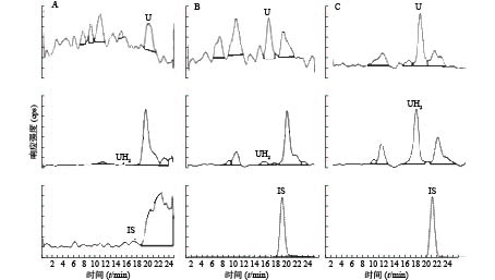
  • U和UH2的出峰时间以及峰型良好,代替血浆经过前处理后,对待测组分的测定没有干扰,内标对分析物的测定也没有干扰,且能很好分离,结果见图1

  • U和UH2的线性范围是10.0~1 500.0 ng/ml,以空白BAS中U和UH2的浓度为横坐标(X),U和UH2与内标化合物氯尿嘧啶的峰面积比为纵坐标(Y),进行最小二乘法加权(权重系数为1/χ2),U和UH2的线性回归方程分别是Y=0.27X+0.0022、Y=0.58X+0.0380,r均>0.990,表明线性关系良好。

  • 取定量下限、低、中、高标准添加血浆样本按照前处理方法进行处理,每个浓度样品平行制备5份进行分析,连续3 d重复操作,根据当天的标准曲线计算当天实测样本浓度,计算样本在低、中和高浓度下的日内、日间精密度和准确度,结果显示,精密度和准确度的偏差均在15%左右。准确度相对偏差在20%范围内时,最低定量下限精密度偏差不大于20%,结果见表1

    分析物标示浓度 (ng/ml)日内日间
    测定浓度 (ng/ml)精密度(CV%)准确性(RE%)测定浓度 (ng/ml)精密度(CV%)准确性(RE%)
    尿嘧啶1010.2±0.383.742.5810.12±0.787.701.18
    2020.63±1.215.873.1519.97±1.356.74−0.16
    500529.73±4.640.885.95484.32±35.727.37−3.12
    10001093.33±25.102.309.331098.25±25.162.299.82
    二氢尿嘧啶1010.32±0.716.863.1810.28±0.656.372.77
    2019.98±2.1910.95−0.1219.86±1.859.31−0.72
    500517.51±10.692.073.50515.66±10.362.013.13
    10001079.83±17.911.667.981080.11±24.502.278.01
  • 取低、高2个浓度的样本进行基质效应和提取回收率考察,结果显示,U、UH2及内标氯尿嘧啶的基质效应和提取回收率良好,结果均较稳定,结果见表2

    分析物标识浓度(ng/ml)基质效应提取回收率
    平均基质效应CV(%)平均回收率CV(%)
    尿嘧啶1000101.006.1594.989.01
    2099.993.63100.017.64
    二氢尿嘧啶100085.722.07106.471.58
    2093.584.5399.549.77
  • 考察低、高2个浓度的血浆样品经历3次冷冻与解冻循环的稳定性、血浆样品在室温(25 ºC)放置6 h后经样品处理后稳定性和血浆样品经样品处理后室温放置24 h的稳定性,结果显示,3次冻融、6 h室温(25 ºC)条件下和24 h放置自动进样器中的稳定性均符合要求,结果见表3

    分析物冻融3次室温放置6 h置自动进样器24 h
    尿嘧啶100.7198.3493.49106.60108.20107.99
    二氢尿嘧啶92.6792.6493.61107.26106.97107.15
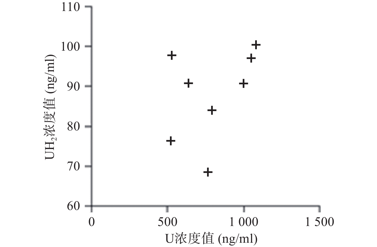
  • 应用本研究所建立的方法,对8名健康成人的血浆样本测定分析,在样本实测过程中, 同时插入已知浓度的随行质控样本(QC样本), 随时监控样本测定的准确度。U和 UH2 浓度测定结果见图2

  • U和UH2是人体内常见的两种物质,且同核酸的代谢密切相关,由于U和UH2均为人体内源性物质,故不能采用人源的基质进行方法学的开发及验证,通过查阅资料,选择了不含U和UH2的3%牛血清蛋白作为基质进行方法学的开发[12]。也有文献报道采用去除U和UH2的人源血浆基质进行方法学实验[9],但基质的来源较珍贵,不适合方法的普及,所以选用3%牛血清蛋白作为替代基质。

  • U因其特殊的化学性质,在大部分的色谱柱上均没有保留。U和UH2的LogP值分别为−0.707和−0.840,有较强的亲水性,决定了其不保留的性质。在定量方法的开发过程中,先后采用了Agilent Zorbax SB-C18色谱柱,Agilent Zorbax Eclipse-C18色谱柱,Waters Atlantis T3色谱柱,Waters Xselect色谱柱,Waters Xbridge等色谱柱来进行条件摸索,但上述色谱柱对U和UH2均没有保留。最后,采用Agilent Zorbax SB-Aq对U和UH2进行定量分析,该色谱柱对强极性的化合物有较好的保留效果,同时,兼容100%的起始流动相也对保留产生了良好的结果。

  • 本研究还分别考察了几种常见的处理方法,包括甲醇和乙腈的蛋白沉淀、Waters Oasis HLB萃取板的固相萃取以及乙酸乙酯,甲基叔丁基醚,二氯甲烷/三氯甲烷,环己烷进行的液液萃取,结果发现乙酸乙酯对U和UH2的萃取效果较好,同时,还分别考察了5%、10%、20%、30%、40%、50%的异丙醇、乙酸乙酯溶液对U和UH2的萃取效果,结果发现,单纯的乙酸乙酯对待测化合物具有较好的提取效率。提取回收率均高于90%,且RSD<10%。另外对3%牛血清蛋白的基质效应进行了考察,结果发现平均基质效应在85%~101%之间,RSD<7%,说明该前处理方法对于基质的清除较为彻底,测定结果稳定,没有明显的基质干扰。

    本研究虽重在方法开发,收集的样本数量较少,但从测定的U和UH2浓度分布来看DPD对内源性U的代谢存在个体差异,建议临床应用5-氟尿嘧啶及其卡培他滨筛查DPD总体活性[12-13],后续可进一步扩大样本数量进行深入研究。

  • 本实验建立了一种快速,稳定,高灵敏度的UHPLC-MS/MS方法,可用于测定人体内源性物质U和UH2的含量,从代谢物的角度评价DPD酶的活性,从而协助临床医生制定化疗药物5-氟尿嘧啶及其口服前药卡培他滨合理的用量,以较低的毒副反应获得最大的临床疗效。

Reference (13)

Catalog

    /

    DownLoad:  Full-Size Img  PowerPoint
    Return
    Return